- Arms
- Right arm IV hectocotylized.
 Click on an image to view larger version & data in a new window
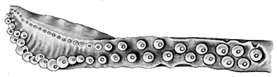
Figure. Oral view of the hectocotylus of L. dislocata, holotype, immature male. Drawing from Young (1972).
- Tentacles
- Medial suckers of club manus about 3X diameter of marginal suckers.
- Alternating suckers and knobs of locking apparatus extend down about half tentacle length; suckers on stalk number 10-13.
Click on an image to view larger version & data in a new window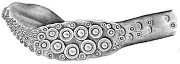
Figure. Oral view of the tentacular club of L. dislocata. Drawing from Young (1972).
- Suckers Click on an image to view larger version & data in a new window
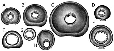
Figure. Representative suckers of L. dislocata. A-C, D - Largest suckers from arms I-IV respectively. The suckers are from the holotype (125 mm GL) which is a male. Suckers from females are smaller and have larger aperatures. E - Largest sucker from arm III of a female (136 mm GL). F - Medial sucker from club manus, holotype. Drawings A-F retain relative sizes. G - Marginal sucker from club manus, holotype. Drawing G has double the enlargement of suckers A-F. H - Enlarged sucker from tip of hectocotylus (137 mm GL). Drawings from Young (1972).
- Mantle
- Mantle cartilagenous strip with second complex tubercule from point of fusion displaced toward the median line.
- Large complex tubercules of strip generally separated by 2 or 3 small tubercules usually with single cusp.
- Complex tubercules in middle of strip with tripartite construction, medial portion with usually 3 cusps aligned in row, lateral sections with more variable arrangement.
- Construction and arrangement of tubercules somewhat variable.
- At nuchal mantle fusion, two small rounded tubercules, one on either side, present at anterior end of cartilagenous pad.
Click on an image to view larger version & data in a new window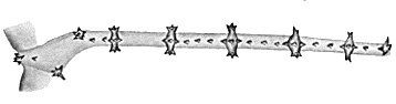
Figure. Ventral view of the left cartilagenous mantle band of L. dislocata showing tubercules, 136 mm GL. Drawing from Young (1972).
Leachia dislocata: Description continued
Richard E. Young and Katharina M. Mangold (1922-2003)About This Page

University of Hawaii, Honolulu, HI, USA
Katharina M. Mangold (1922-2003)
Laboratoire Arago, Banyuls-Sur-Mer, France
Page copyright © 1998 and Katharina M. Mangold (1922-2003)
 Page: Tree of Life
Leachia dislocata: Description continued
Authored by
Richard E. Young and Katharina M. Mangold (1922-2003).
The TEXT of this page is licensed under the
Creative Commons Attribution-NonCommercial License - Version 3.0. Note that images and other media
featured on this page are each governed by their own license, and they may or may not be available
for reuse. Click on an image or a media link to access the media data window, which provides the
relevant licensing information. For the general terms and conditions of ToL material reuse and
redistribution, please see the Tree of Life Copyright
Policies.
Page: Tree of Life
Leachia dislocata: Description continued
Authored by
Richard E. Young and Katharina M. Mangold (1922-2003).
The TEXT of this page is licensed under the
Creative Commons Attribution-NonCommercial License - Version 3.0. Note that images and other media
featured on this page are each governed by their own license, and they may or may not be available
for reuse. Click on an image or a media link to access the media data window, which provides the
relevant licensing information. For the general terms and conditions of ToL material reuse and
redistribution, please see the Tree of Life Copyright
Policies.


