


Leachia
Richard E. Young, Katharina M. Mangold (1922-2003), and Michael Vecchione
Six of the 14 nominal species of Leachia are considered valid by Voss, et al. (1992). These authors suggest that 11 species exist in the genus, 5 of which are undescribed.
- Leachia atlantica
- Leachia cyclura
- Leachia danae (Joubin, 1931)
- Leachia dislocata Young, 1972
- Leachia lemur
- Leachia pacifica
- Leachia sp. A
Introduction
Species of Leachia are easily recognized by the straight cartilagenous, tubercular strip on the mantle that arises from each point of funnel-mantle fusion, the slender, tapering mantle and the terminal fins with a combined oval shape.
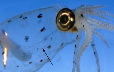
Figure. Ventrolateral view of the antrior region of Leachia dislocata showing a transparent, cartilagenous strip bearing tubercules (arrow). Tubercules can be difficult to see because of their transparency. Photograph by R. Seapy.
Leachia species, however, are not easily recognized. Many names exist in the literature and most have poor descriptions associated with them and are based on paralarvae. At present, geographical location is one of the most important characteristics used in identification. The type localities of the named species are:
Atlantic Ocean
- Leachia atlantica - eastern North Atlantic, 36°, 10°W.
- Leachia lemur - western North Atlantic, east of Cape Hatteras, 35°N, 73°W,.
- Leachia sp. A - western South Atlantic, 39°S, 53°W.
Indian Ocean
- Leachia cyclura - south Indian Ocean, 37°S 33°E.
Pacific Ocean
- Leachia danae - (Gulf of Panama).
- Leachia dislocata - off Southern California, eastern temperate Pacific.
- Leachia pacifica - south Pacific Ocean, 15°S, 168°W
Brief diagnosis:
A cranchiin...
- with one cartilagenous strip bearing tubercules on the mantle originating at each point of funnel-mantle fusion.
Characteristics
- Tentacles
- Median suckers of tentacular club greatly enlarged.**
Click on an image to view larger version & data in a new window
Figure. Oral view of the tentacular club of Leachia dislocata, 136 mm ML. Drawing from Young (1972).
- Median suckers of tentacular club greatly enlarged.**
- Head
- Eyes stalked in paralarvae.
- Beaks: Descriptions of L. danae beaks can be found here: Lower beak; upper beak.
- Funnel
- Funnel valve absent.
- Funnel organ: dorsal pad U-shaped with 3-7 papillae.
- Mantle
- Single tuberculate cartilagenous strip on mantle originates at each funnel-mantle fusion.*
- Single tuberculate cartilagenous strip on mantle originates at each funnel-mantle fusion.*
- Fins
- Fins barely unite posterior to gladius (transversely elliptical in combined outline).
- Fins barely unite posterior to gladius (transversely elliptical in combined outline).
- Photophores
- Each eye with 5-21 oval photophores depending on species.
- Photophores on tips of arms III in mature or nearly mature females.
**Unique feature in family where suckers unmodified (not hooks or hook-like suckers)
Comments
This is the only genus in the subfamily Cranchiinae with paralarvae that have stalked eyes. Characteristics are from Voss (1980).
Comparison of species
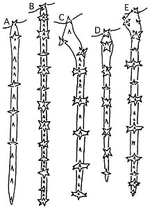
Figure. Comparison of the structure of the mantle tubercular strips among five of the species. A - Leachia lemur, north Sargasso Sea, 42 mm mL. B - Leachia atlantica, subtropical North Atlantic, 54 mm ML. C - Leachia dislocata, off California, 34 mm ML. D - Leachia danae, eastern tropical Pacific, 53 mm ML. E - Leachia pacifica, off Hawaii, 41 mm ML. Drawings from Voss, et al. (1992, p. 191).
Life History
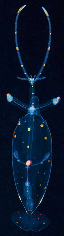
Figure. Dorsal view of L. pacifica, paralarva, 15-25 mm ML (guess). Insitu photograph taken while scuba diving, Hawaii. Note the tubercular strips seen through the mantle and the very long eye stalks. © 2015 Jeffrey Milisen
The paralarvae of the genus (below, left) have a very distinctive appearance. A paralarva has a very long, slender brachial pillar and a pointed tip to the gladius. Paralarvae of Leachia were originally placed in the genus Pyrgopsis and are now often referred to as pyrgopsis paralarvae. The maturing adult (below, right) may, depending on the species, may retain partially stalked eyes as in L. atlantica.
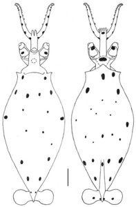
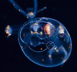
Figure. Left - Dorsal and ventral views of paralarval L. pacifica, 7.9 mm ML, Hawaiian waters. Drawing by R. Young. Right - Anterior view of L. pacifica, 15-25 mm ML (guess), same specimen as seen at the right. The head and arms are out of focus; but the mantle margin is sharp. A circle has been placed over the left mantle-attachment site. Within the circle four anterior tubercules of the tubercular strip are visible including one "displaced" tubercule, a characteristic of this species (as well as L. dislocata found off Southern California). © 2015 Jeffrey Milisen
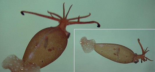
Figure. Posterodorsal and dorsolateral views of the same maturing L. atlantica. The approach to maturity is indicated by the darkly pigmented, terminal arm III photophores and the heavy skin pigmentation. Note that in the lower-right image that a row of ventral-mantle tubercules can be seen along the lower edge of the mantle. Photographs from an ROV video operated from the R/V OKEANOS EXPLORER and taken at a depth of about 1700 in the northern Gulf of Mexico; provided by NOAA's Ocean Exploration program.
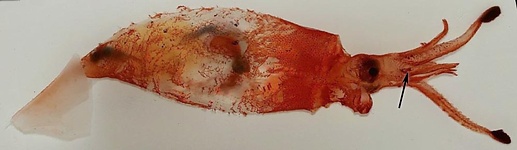
Figure. L. atlantica, 90 mm ML, female, northeast Atlantic. Side view. Arrow points to the remnant of a tentacle presumably lost in situ as maturity approached. The light yellowish material in the posterior third of the mantle may be the developing ovary. Photographed fresh aboard ship after capture in a trawl by M. Vecchione.
Distribution
Vertical distribution
Off Hawaii, L. pacifica has a peculiar vertical distribution pattern which may prove to be common within the genus. Small squid are found in near-surface waters. As sexual maturity approaches, the squid undergoes an abrupt ontogenetic descent. At depths greater than 1000 m males and females become mature. Large photophores develop on the tips of the third arms of females and these are, presumably, used to attract males at great depths where the risk of predation is low.

Figure. Vertical distribution chart of L. pacifica, Hawaiian waters. Captures were made with both open and opening/closing trawls. Bars - Fishing depth-range of opening/closing trawl. Circle - Modal fishing depth for either trawl. Blue color - Night captures. Yellow color - Day captures. Chart modified from Young (1978).
References
Voss, N. A. 1980. A generic revision of the Cranchiidae (Cephalopoda; Oegopsida). Bull. Mar. Sci. 30: 365-412.
Voss N. A., S. J. Stephen and Zh. Dong 1992. Family Cranchiidae Prosch, 1849. Smithson. Contr. Zool., 513: 187-210.
Young, R. E. 1972. The systematics and areal distribution of pelagic cephalopods from the seas off Southern California. Smithson. Contr. Zool. 97:1-159.
Young, R. E. 1978. Vertical distribution and photosensitive vesicles of pelagic cephalopods from Hawaiian waters. Fish. Bull. 76: 583-615.
Title Illustrations

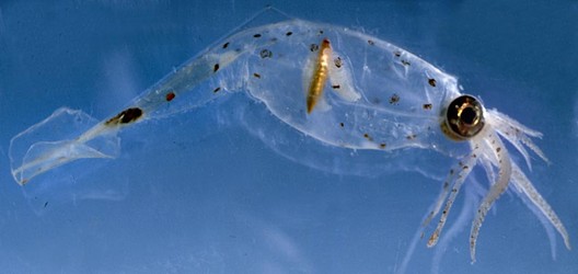
| Scientific Name | Leachia dislocata |
|---|---|
| Location | Off Southern California |
| Specimen Condition | Live Specimen |
| View | Side |
| Size | 100 mm ML (estimate) |
| Image Use |
 This media file is licensed under the Creative Commons Attribution-NonCommercial License - Version 3.0. This media file is licensed under the Creative Commons Attribution-NonCommercial License - Version 3.0.
|
| Copyright |
©

|

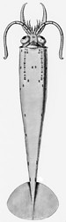
| Scientific Name | Leachia dislocata |
|---|---|
| Location | Off Southern California |
| Reference | from Young, R. E. 1972. The systematics and areal distribution of pelagic cephalopods from the seas off Southern California. Smithson. Contr. Zool. 97:1-159. |
| Acknowledgements | Drawing by Constance McSweeny. |
| View | Ventral |
| Size | 110 mm ML |
| Image Use |
This media file is licensed under the Creative Commons Attribution-NonCommercial License - Version 3.0.
|
| Copyright |
©
|
About This Page
University of Hawaii, Honolulu, HI, USA
Katharina M. Mangold (1922-2003)
Laboratoire Arago, Banyuls-Sur-Mer, France
National Museum of Natural History, Washington, D. C. , USA
Page copyright © 2018 , Katharina M. Mangold (1922-2003), and
 Page: Tree of Life
Leachia .
Authored by
Richard E. Young, Katharina M. Mangold (1922-2003), and Michael Vecchione.
The TEXT of this page is licensed under the
Creative Commons Attribution-NonCommercial License - Version 3.0. Note that images and other media
featured on this page are each governed by their own license, and they may or may not be available
for reuse. Click on an image or a media link to access the media data window, which provides the
relevant licensing information. For the general terms and conditions of ToL material reuse and
redistribution, please see the Tree of Life Copyright
Policies.
Page: Tree of Life
Leachia .
Authored by
Richard E. Young, Katharina M. Mangold (1922-2003), and Michael Vecchione.
The TEXT of this page is licensed under the
Creative Commons Attribution-NonCommercial License - Version 3.0. Note that images and other media
featured on this page are each governed by their own license, and they may or may not be available
for reuse. Click on an image or a media link to access the media data window, which provides the
relevant licensing information. For the general terms and conditions of ToL material reuse and
redistribution, please see the Tree of Life Copyright
Policies.
- Content changed 29 March 2018
Citing this page:
Young, Richard E., Katharina M. Mangold (1922-2003), and Michael Vecchione. 2018. Leachia . Version 29 March 2018 (under construction). http://tolweb.org/Leachia/19544/2018.03.29 in The Tree of Life Web Project, http://tolweb.org/


