


Leachia danae
Henk-Jan Hoving, Stephanie Bush, and Richard E. Young
Introduction
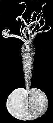
Figure. Ventral view of L. danae,
male, ca. 155 mm ML.
Drawing from Joubin, 1931.
L. danae is probably the largest member of the genus. Females reach a length of, at least, 183 mm ML.
Characteristics
- Arms
- Arms attenuate with more than 100 suckers (fide Joubin, 1931).
- Males with arms III subequal in length with arm I and II (fide Joubin, 1931, see illustration at right).
- Male hectocotylus a flag-like structure at the distal end of right arm IV.
Click on an image to view larger version & data in a new window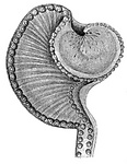
Figure. Hectocotylized tip of the right arm III of a mature male
of L. danae, ca 155 mm ML. Drawing from Joubin, 1931.
- Arms I, II, IV with smooth sucker rings.
- Suckers on mid-arm III of females each with greatly enlarged, hook-like tooth.
Click on an image to view larger version & data in a new window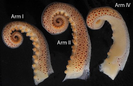
Figure. Side views of arms I, II and IV of L. danae, female, 179 mm ML.
Photographs by Henk-Jan Hoving.Click on an image to view larger version & data in a new window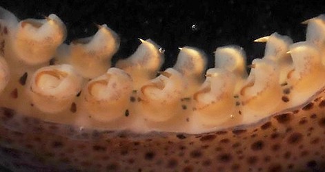
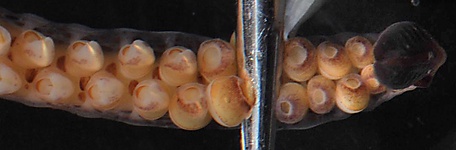
Figure. Arm III of L. danae, female, 179 mm ML. Top - Oral-lateral view, mid-arm, showing large, hook-like tooth on each sucker. Note the small, slender secondary cusps visible on some of the sucker rings. Bottom - Oral view, distal arm, showing transformation of suckers with hooks into simple globular suckers near arm tip. Note black arm-tip photophores curved toward viewer. Silver bar is a pair of forceps. Photographs by Henk-Jan Hoving.
- Tentacle
- Greatly reduced in near-mature female; suggesting resorption rather than autotomy of the subadult tentacle.
Click on an image to view larger version & data in a new window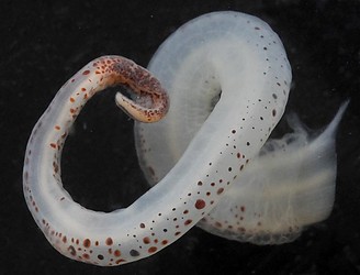
Figure. Right tentacle remnant of the near mature female of L. danae, 179 mm ML. Note the weak musculature and the heavily pigmented club with traces of the protective membranes. Photograph by Henk-Jan Hoving.
- Greatly reduced in near-mature female; suggesting resorption rather than autotomy of the subadult tentacle.
- Head
- Beaks: Descriptions can be found here: Lower beak; upper beak.
- Beaks: Descriptions can be found here: Lower beak; upper beak.
- Photophores
- 20 small, "round" photophores present on eyes of adult: 5 on ventrolateral face (no. 1 and 5 larger that 2-4); 7 on dorsolateral face (all small but no. 6 and 12 larger than 7-11); series of 8 (nos. 13-20) running from anterior to posterior edges of eyeball while passing along the medioventral surface of the eyeball (photophore no. 20 on posterior margin rises well off surface of eyeball.) Click on an image to view larger version & data in a new window
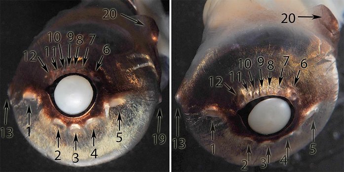
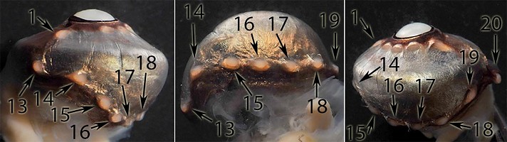
Figure. Ocular photophores of L. danae, female, 179 mm ML. Top left - Frontal view. Top right - Dorsofrontal view. Bottom left - Anterior view. Bottom middle - Ventromedial view. Bottom right - Posteroventral view. Photographs by Henk-Jan Hoving.
- Photophores on tips of all arms in maturing females.
Click on an image to view larger version & data in a new window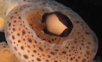
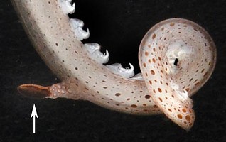
Figure. Oral view of the terminal arm I photophore of L. danae, 179 mm ML. Left - Preserved squid showing the light-colored photogenetic surface and the dark brown, only slightly expanded lids that can conceal light emission. Photograph by Henk-Jan Hoving. Right - Living squid showing the distal portion of arm III coiled into a near figure-8 with the terminal photophore just visible (arrow) and fully closed. Photograph by Steven Haddock.
- 20 small, "round" photophores present on eyes of adult: 5 on ventrolateral face (no. 1 and 5 larger that 2-4); 7 on dorsolateral face (all small but no. 6 and 12 larger than 7-11); series of 8 (nos. 13-20) running from anterior to posterior edges of eyeball while passing along the medioventral surface of the eyeball (photophore no. 20 on posterior margin rises well off surface of eyeball.)
- Reproductive structures
- Viscera.
Click on an image to view larger version & data in a new window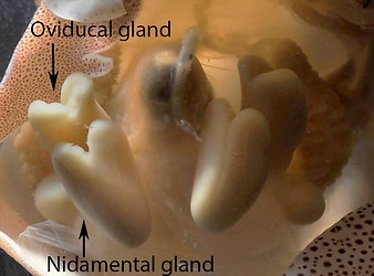
Figure. Ventral view of the viscera of L. danae, 179 mm ML showing the strong similarity between oviducal and nidamental glands. Photograph by Henk-Jan Hoving.
- Peculiar structures: Tissue surrounding the gladius and material looking like tiny "cotton-balls" above the gladius and on the posterodorsal head. The photographs below compare the tissue over the gladius in the living squid and in the preserved squid like forming flaps anteriorly forms a trough dorsal to the glades. This tissue, along with the gladius is covered by the dorsal skin. In the trough, external to the skin, are tiny "cotton balls" which are also found on the posterdorsal surface of the head.
We have no direct evidence regarding the nature or function of the "cotton balls." Our best guess is that they represent glandular structures that function in sexual attraction.
Click on an image to view larger version & data in a new window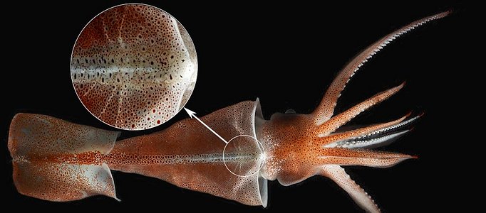
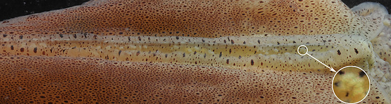
Figure. Dorsal view of the mantle mid-region of the female L. danae, 179 mm ML, showing the unusual structures associated with the gladius. Top - Living squid in a shipboard tank. The insert shows a higher magnification of this structure. The radiating white lines are, presumably, mantle nerves arising from the stellate ganglia. Photograph by Steven Haddock. Bottom - Preserved squid. There now appears to be a longitudinal trough in the structure suggesting that it can be opened or closed in the living squid. Note that the tiny "cotton-balls" have a somewhat granular appearance (see insert). Photograph by Henk-Jan Hoving.
- Viscera.
Life History
Young L. danae (left) and near-adult (right):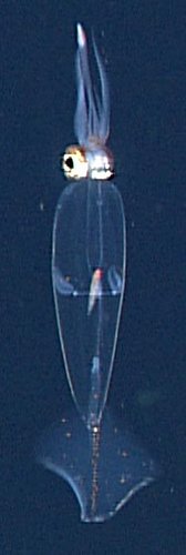
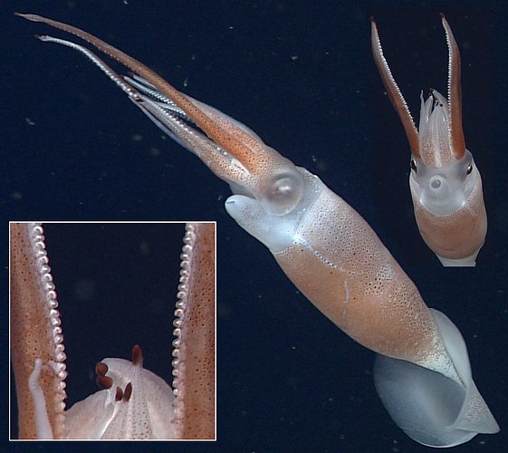
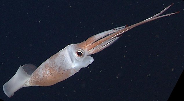
Figure. Left - Young L. danae dorsal view, In situ ROV photographs at 406 m depth. Note the oval fin shape characteriestic of the genus. Right - Various views of L. danae, near-adult, 179 mm ML. In situ ROV photographs at 1368 m depth. Left insert - Close-up view showing the arm-tip photophores, the tip of the regressed right tentacle, and the hook-like suckers of arms III. Middle - Side view showing squid with eye shut: The iris is nearly closed, leaving only a small circular pupil at the anteroventral edge of the covered lens. Also note the left tentacle stub. Right insert - Anteroventral view of the same squid. Note the regressed right tentacle. The squid was jetting backwards with fins folded against the body. © 2013 MBARI .
Distribution
Type locality: Eastern Tropical Pacific off Panama at 6° 40'N, 80°47'W. Known also from off Hawaii and the Gulf of California.Title Illustrations

| Scientific Name | Leachia danae |
|---|---|
| Location | Gulf of California at 24.18°N, 109.63°W |
| Comments | In situ photograph of L. danae taken at a depth of 1368 m. |
| Acknowledgements | Image courtesy of the Monterey Bay Aquarium Research Institute (MBARI). You must obtain permission from MBARI to use this photo; please contact pressroom@mbari.org for further information |
| Specimen Condition | Live Specimen |
| Identified By | R. E. Young |
| Sex | Female |
| Life Cycle Stage | Nearly mature |
| View | Right-side |
| Size | 179 mm ML |
| Copyright | © 2013 MBARI |
About This Page

GEOMAR-Helmholtz Centre for Ocean Research Kiel
Monterey Bay Aquarium Research Institute
University of Hawaii, Honolulu, HI, USA
Page copyright © 2018 , , and
 Page: Tree of Life
Leachia danae .
Authored by
Henk-Jan Hoving, Stephanie Bush, and Richard E. Young.
The TEXT of this page is licensed under the
Creative Commons Attribution-NonCommercial License - Version 3.0. Note that images and other media
featured on this page are each governed by their own license, and they may or may not be available
for reuse. Click on an image or a media link to access the media data window, which provides the
relevant licensing information. For the general terms and conditions of ToL material reuse and
redistribution, please see the Tree of Life Copyright
Policies.
Page: Tree of Life
Leachia danae .
Authored by
Henk-Jan Hoving, Stephanie Bush, and Richard E. Young.
The TEXT of this page is licensed under the
Creative Commons Attribution-NonCommercial License - Version 3.0. Note that images and other media
featured on this page are each governed by their own license, and they may or may not be available
for reuse. Click on an image or a media link to access the media data window, which provides the
relevant licensing information. For the general terms and conditions of ToL material reuse and
redistribution, please see the Tree of Life Copyright
Policies.
- First online 21 January 2014
- Content changed 21 January 2014
Citing this page:
Hoving, Henk-Jan, Stephanie Bush, and Richard E. Young. 2014. Leachia danae . Version 21 January 2014 (under construction). http://tolweb.org/Leachia_danae/19587/2014.01.21 in The Tree of Life Web Project, http://tolweb.org/


