


Babesiidae
Jan Votýpka


This tree diagram shows the relationships between several groups of organisms.
The root of the current tree connects the organisms featured in this tree to their containing group and the rest of the Tree of Life. The basal branching point in the tree represents the ancestor of the other groups in the tree. This ancestor diversified over time into several descendent subgroups, which are represented as internal nodes and terminal taxa to the right.
You can click on the root to travel down the Tree of Life all the way to the root of all Life, and you can click on the names of descendent subgroups to travel up the Tree of Life all the way to individual species.
For more information on ToL tree formatting, please see Interpreting the Tree or Classification. To learn more about phylogenetic trees, please visit our Phylogenetic Biology pages.
close boxIntroduction
Family Babesiidae Poche, 1913
Relatively large, piriform, round or oval parasites with the apical complex reduced to a polar ring, rhoptries and subpellicular microtubules. Conoid is present only in some species. At least some stages are found in erythrocytes. Trophozoites dividing by binary fission usually produce two (rarely four) daughter merozoites, which enter new red blood cells. Other stages are in lymphocytes, histiocytes, erythroblasts and other blood cells. Vertebrate hosts are mammals and birds, vectors are ticks where binary fission and schizogony occur, but there is probably no sexual reproduction.
Characteristics
Development in mammalian erythrocytes is relatively well documented. However, the development within the invertebrate host is quite complicated and known only for a few species. After ingestion by a tick, the parasites leave the blood cell to develop into a spiked gamete (ray bodies), which fuse into a motile zygote and form a primary kinete. Due to the penetration of the elongated kinetes (vermicules) into numerous internal organs of the tick, including the ovaries, the infection can be passed through transovarial transmission into the next generation, with the ticks remaining infectious for several generations. Further development occurs in the hemocoel and various organs, where Babesia produces new secondary kinetes, some of which migrate into the salivary glands. During blood feeding that takes several hours or days, piroplasms rapidly multiply and eventually transform into sporonts and infectious sporozoites. Finally, sporozoites from the tick saliva are injected into the vertebrate host, where they directly infect red blood cells and develop into the well-known stages.
The first piroplasmid was described from the blood of cattle during the 1880s epidemic of Texas cattle fever in the United States. Similar blood stages, later included in the genus Babesia, were described in 1888 by Victor Babeş. Only a few years later, Theobald Smith and Frank Kilbourne successfully transmitted a related organism to non-infected cattle via a tick, and showed for the first time that invertebrates can serve as vectors of a parasitic disease.
Title Illustrations
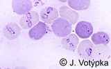
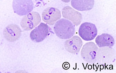
| Scientific Name | Babesia rodhaini |
|---|---|
| Location | Parasites in blood of mice |
| Image Use |
 This media file is licensed under the Creative Commons Attribution-NonCommercial License - Version 3.0. This media file is licensed under the Creative Commons Attribution-NonCommercial License - Version 3.0.
|
| Copyright |
© 2008 Jan Votýpka

|

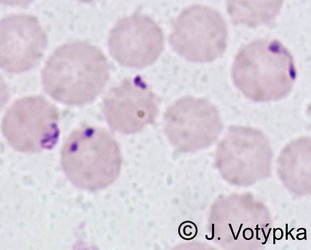
| Scientific Name | Babesia divergens |
|---|---|
| Location | Parasites in blood of cattle |
| Image Use |
This media file is licensed under the Creative Commons Attribution-NonCommercial License - Version 3.0.
|
| Copyright |
© 2008 Jan Votýpka
|
About This Page
This page is being developed as part of the Tree of Life Web Project Protist Diversity Workshop, co-sponsored by the Canadian Institute for Advanced Research (CIFAR) program in Integrated Microbial Biodiversity and the Tula Foundation.
Jan Votýpka
Department of Parasitology, Charles University
Correspondence regarding this page should be directed to Jan Votýpka at
vapid@natur.cuni.cz
Page copyright © 2011 Jan Votýpka
 Page: Tree of Life
Babesiidae.
Authored by
Jan Votýpka.
The TEXT of this page is licensed under the
Creative Commons Attribution-NonCommercial License - Version 3.0. Note that images and other media
featured on this page are each governed by their own license, and they may or may not be available
for reuse. Click on an image or a media link to access the media data window, which provides the
relevant licensing information. For the general terms and conditions of ToL material reuse and
redistribution, please see the Tree of Life Copyright
Policies.
Page: Tree of Life
Babesiidae.
Authored by
Jan Votýpka.
The TEXT of this page is licensed under the
Creative Commons Attribution-NonCommercial License - Version 3.0. Note that images and other media
featured on this page are each governed by their own license, and they may or may not be available
for reuse. Click on an image or a media link to access the media data window, which provides the
relevant licensing information. For the general terms and conditions of ToL material reuse and
redistribution, please see the Tree of Life Copyright
Policies.
- First online 18 May 2011
- Content changed 18 May 2011
Citing this page:
Votýpka, Jan. 2011. Babesiidae. Version 18 May 2011 (under construction). http://tolweb.org/Babesiidae/68076/2011.05.18 in The Tree of Life Web Project, http://tolweb.org/

