

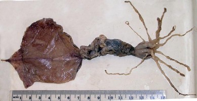
Figure. Top - Dorsal view of the subadult M. atlantica, 72 mm ML. Scale bar is 1 cm. Bottom - Ventral view of the adult M. atlantica, 127 mm ML. Photographs by M. Vecchione.
Introduction
We describe here two male specimens from the Gulf of Mexico which we place in M. atlantica. The taxonomy of the Atlantic species of Magnapinna contains considerable uncertainty, and our placement of the specimens here is tentative. Most of the following features have been taken from the smaller, subadult male which is in reasonably good condition. The larger, mature male is in very poor condition and information from it is mostly limited to the male reproductive system and the beaks.
Characteristics
Description
- Arms
- Suckers on proximal-arm in two series and closely packed.
- Proximal-arm suckers with smooth inner rings; fleshy diameter of large suckers about 0.6 mm; diameter of outer ring about 0.3 mm; sucker orifice diameter about 40% of maximum diameter of inner ring.
- Transition between proximal- and distal-arms marked by abrupt changeover from large proximal-arm suckers to minute distal-arm suckers (often one or two suckers of intermediate size present); diameter of distal-arm suckers about 0.1 mm initially, shortly grading to about 0.06-0.08 mm.
- Distal-arms, apparently, with slightly narrower diameter than ends of proximal-arms and may have less pigment.
Click on an image to view larger version & data in a new window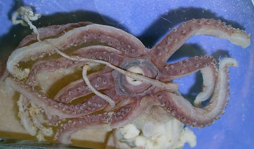
Figure. Oral view of the brachial crown of M. atlantica, 72 mm ML, showing arrangement of proximal-arm suckers. Photograph by M. Vecchione.
Click on an image to view larger version & data in a new window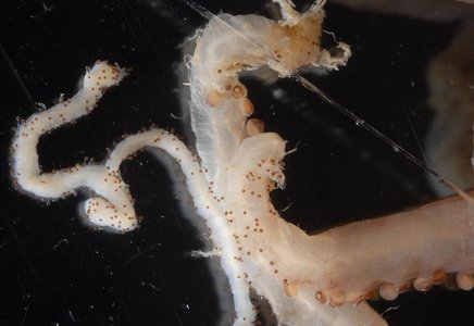
Figure. Side views of two proximal-arms of M. atlantica showing transition to distal-arms. Adjacent to the arms on the left is a portion of a distal-tentacle, 72 mm ML. Compare sucker sizes on distal-arms and tentacle. Photograph by R. Young.
Click on an image to view larger version & data in a new window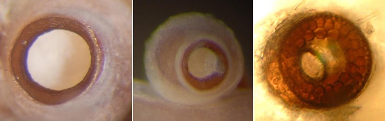
Figure. Arms and tentacle suckers of M. atlantica. Left - Oral view of the outer ring of a large proximal-arm sucker, 127 mm ML. Middle - Oral view of a large proximal-arm sucker, 72 mm ML. Note the small sucker orifice. Rght - Oral view of a distal-arm sucker. Photographs by R. Young.
- Tentacle
- Proximal-tentacles long, thin, cylindrical and taper rapidly; thickness at base much thinner than the arms at their bases. The proximal-tentacle appears to grade into the distal tentacle without a clear separation between them. We use the first minute sucker and the last chromatophore to identify the changeover.
- Distal-tentacle with numberous, small suckers about 0.06-0.08 mm in diameter; suckers apparently irregularly arranged and cover much of tentacle circumference although a bare strip appears to be present along one surface.
- Distal-tentacle suckers with smooth inner rings.
Click on an image to view larger version & data in a new window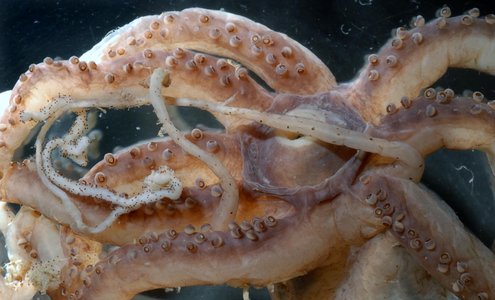
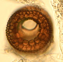
Figure. Left - Oral view of the brachial crown of M. atlantica FMNH 308252, showing tentacles. This image is similar to the photograph at the top of this section but has higher magnification.The top arrow points the the glandular region, the second arrow points to the first clearly recognizable sucker. More proximal suckers are possible but damage prevents resolving this. However, chromatophores can be detected on the tentacle to within about 1 mm of the first sucker and no chromatophores can be recognized distal to this point. Right - Oral view of a distal-arm sucker, same specimen. Photographs by R. Young.
Click on an image to view larger version & data in a new window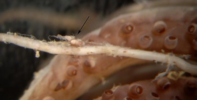
Figure. View of the glandular structures (arrow) on the proximal-tentacle of M. atlantica. Note that the glands are barely attached to the tentacle. Photographs by R. Young.
Click on an image to view larger version & data in a new window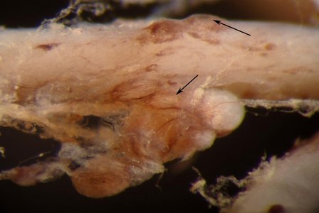
Figure. Close view of the glandular structures on the proximal-tentacle of M. atlantica. Arrows point to apparent openings from glands to the exterior. Most of the glands are the large, spherical structures dangling below the bottom arrow. Photograph by R. Young.
- Proximal-tentacles long, thin, cylindrical and taper rapidly; thickness at base much thinner than the arms at their bases. The proximal-tentacle appears to grade into the distal tentacle without a clear separation between them. We use the first minute sucker and the last chromatophore to identify the changeover.
- Head
- Eyes large.
- Beaks. Description of the peculiar beaks of M. atlantica can be found here.
- Radula. Radula with tricuspid rhachidian teeth and bicuspid first lateral teeth. Marginal plates barely detectable.
Click on an image to view larger version & data in a new window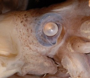
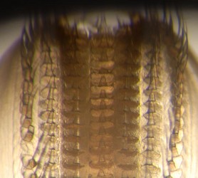
Figure. Left - Side view of the head of M. atlantica showing the slightly damaged eye, 72 mm ML. Right - Radula of M. atlantica, 127 mm ML. Photograph by R. Young.
- Funnel
- Funnel locking-apparatus with oval depression.
- Dorsal funnel organ swollen and covered with debris; exact shape not determined. Ventral pads elongate, triangular-shaped with base of triangle at posterior end.
- Small, weak, funnel valve present.
Click on an image to view larger version & data in a new window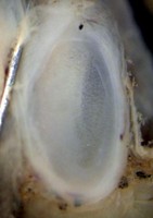
Figure. Frontal view of the funnel component of the funnel/mantle locking apparatus of M. atlantica FMNH 308253. Photograph by M. Vecchione.
- Fins
- Fins about 75% of ML; slightly broader than long.
- Short tail present but gladius broken off.
- Apex angle of fins nearly 90°????.
- Fins about 75% of ML; slightly broader than long.
- Pigmentation
- Epithelial pigmentation, darker on oral surfaces, covers squid except possibly on distal-tentacles (initial portion, at least, of distal-arms has some epithelial pigment); scattered chromatophores overlie epithelial pigmentation on most surfaces (including proximal-tentacles) except oral surfaces (and probably aboral surfaces) of arms, buccal membrane and outer lips. Chromatophores not recognized on mature male but skin badly damaged.
Click on an image to view larger version & data in a new window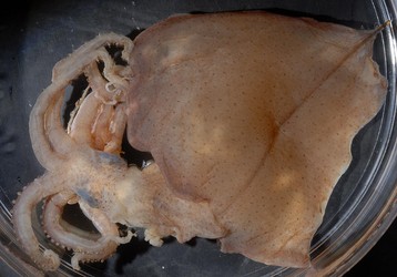
Figure. Left - Dorsal view of M. atlantica, 72 mm ML, showing pigmentation. Right - Dorsal view of a region of the fin near its edge, 127 mm ML, showing absence of chromatophores and what appear to be tiny vesicles. Photographs by R. Young.
- Epithelial pigmentation, darker on oral surfaces, covers squid except possibly on distal-tentacles (initial portion, at least, of distal-arms has some epithelial pigment); scattered chromatophores overlie epithelial pigmentation on most surfaces (including proximal-tentacles) except oral surfaces (and probably aboral surfaces) of arms, buccal membrane and outer lips. Chromatophores not recognized on mature male but skin badly damaged.
- Gladius
- Gladius not examined.
- Gladius not examined.
- Viscera
- Viscera anteriorlly located.
- Anal valves and small ink-sac present.
- Spermatophore with very short ejaculatory apparatus, long, coiled cement gland and uncoiled sperm mass.
Click on an image to view larger version & data in a new window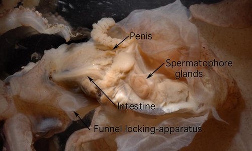
Figure. Ventral view of viscera within the mantle cavity of M. atlantica, 72 mm ML. Photograph by R. young.
Click on an image to view larger version & data in a new window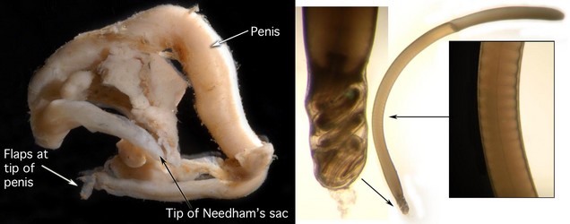
Figure. Left - Penis and portions of the testis and spermatophore glands of M. atlantica, 127 mm ML, that had been torn free of the viscera apparently during capture. Right - Spermatophore of M. atlantica that we removed from the end of the Needham's sac. No other spermatophores were seen but nothing more was dissected. Left figure is an enlargement of the ejaculatory apparatus. Right figure is an enlargement of a portion of the cement body. Photographs by R. Young.
- Measurements and counts
*Length of mantle anterior to fin attachment.Holotype Paratype FMNH 308252 FMNH 308253 Sex - - Immature male Mature male Mantle length 59 53 72 127 Tail length 4+ 1+ 3+ 6+ Mantle width 9 8 13 17 Muscular mantle length 23 17 38 66 Free-mantle length*
12 8 26 44 Fin length
53 41 54 83+ Fin width 60 ca. 52 68 94 Head width 8 7 11 - Head length 6 7 8
21 Eye diameter 4 4 5 - Proximal-arm I length 11.5 8.8 26 - Proximal-arm II length
12 9.3 25 - Proximal-arm III length
11.5 8.8 20 - Proximal-arm IV length
12.5 10 21 - Sucker counts, Prox.-arm I - - 39
- Sucker counts, Prox.-arm II
44 - 38+
- Sucker counts, Prox.-arm III
- 37 41 - Sucker counts, Prox.-arm IV
- - 43 - Proximal-tentacle length 10 / 7 6 / 7 36 / - -
Comments
We assume that these two squid belong to the same species, although the larger specimen is badly damaged, since they look superficially similar and were taken at nearly the same date from nearly the same location.
M. atlantica is separated from M. talismani and Magnapinna sp. C most clearly by the presence of glandular structures on the proximal-tentacles. We show here that these structures apparently undergo a relative decrease with size as the squid grows and that they can be easily lost. Therefore, the absence of the glandular structures in other species with narrow-proximal tentacles is now questionable. Magnapinna sp. B (95 mm ML) has a deep reddish epithelial pigmentation without detectable chromatophores. The larger specimen described here (127 mm ML) also has no detectable chromatophores but was badly damaged. Magnapinna sp. B also seems to have broader apperatures on their large arm suckers. Magnapinna sp. C (79 mm ML) is comparable to the smaller squid described here (72 mm ML) has chromatophores but no epithelial pigmentation. However it has been in preservation for over 50 years. This specimen needs to be re-examined.


