
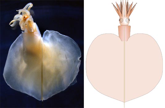
Figure. Dorsal views of M. atlantica, holotype. Left - Photograph of preserved squid. Right - Drawing. Images from Vecchione and Young (submitted), modified.
- Arms
- Some proximal-arms have numerous, small, irregularly arranged suckers near tips but proximal to distal-arms.
- Large suckers of proximal-arms 0.4 mm in diameter; suckers of distal-arms 0.1 mm in diameter (fleshy widths), outer ring just slightly smaller.
- Proximal-arm suckers with smooth inner rings.
- Proximal-arms without protective membranes or keels except arms IV which have broad lateral membranes.
- Tentacles
- Distal-tentacles appear to have multiple series of small suckers.
- Head
- Eye large, eye opening circular and without ocular sinus; pigmented iris present.
- Olfactory organs on long stalks at posterolateral margins of eyes.
- Head without excavation on ventral surface between funnel adductors.
- Beaks not examined.
- Funnel
- Funnel component of the funnel/mantle-locking apparatus approximately with deep, hemispherical groove (see figure below).
- Mantle component of the locking-apparatus nearly semicircular in lateral profile (see figure below), slightly more compressed in anterior profile.
- Mantle
- Mantle wall thick but mostly occupied by gelatinous tissue.
- Fins
- Fins terminal.
- Photophores
- Photophores absent.
- Pigmentation
- Tentacles with numerous chromatophores in glandular region only few, scattered more proximally.
- Arms with few scattered chromatophores aborally at bases otherwise without chromatophores.
- Arms with very light (barely detectable), brown integumental pigmentation at oral bases.
- Gladius
- Gladius not extracted.
- Viscera
- Ink sac, anal valves present; thin, greatly expanded caecum present.
- Cyclindrical, orange digestive gland abuts cephalic cartilage.
- Gonad appears to be an ovary due to its irregular surface; no nidamental glands apparent.
- Measurements
- ML - 59 mm, MW - 9 mm, FL - 53 mm, FW - 60 mm, Tail Length - 4+, Eye diameter - 4 mm, Lens Diameter - 1.8 mm, HL - 6 mm, HW - 8 mm, Arm I (proximal region) 11.5, Arm II (prox. reg.) - 12 mm, Arm III (prox. reg.) - 11.5, Proximal-arm IV - 12.5 mm, TL (left, prox.-tent.) - 10 mm, TL (right, prox.-tent.) - 7 mm, Arm Sucker Count (arm II, left) - 22 pairs of large suckers, Tentacle width at base - 1.4 mm, Arm IV width at base - 2.2 mm.
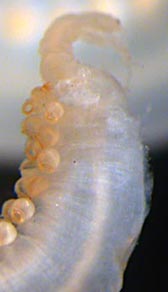
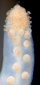
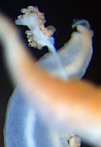
Figure. Arms of M. atlantica, holotype. Left - lateral view of left proximal-arm II with a remnant of the distal-arm. Middle - Oral view of right proximal-arm IV with many small suckers at the tip, but with the distal-arm missing. Right - Aboral view of right arm II showing the nearly-detached basal region of the distal-tentacle bearing small suckers. Photographs from Vecchione and Young (2006), modified.
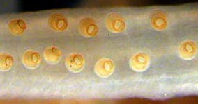
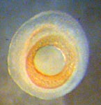
Figure. Oral view of an arm (left) and a sucker (right) of M. atlantica, holotype. Note the lack of arm protective membranes. Photographs by R. Young.
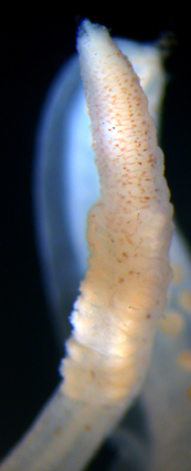
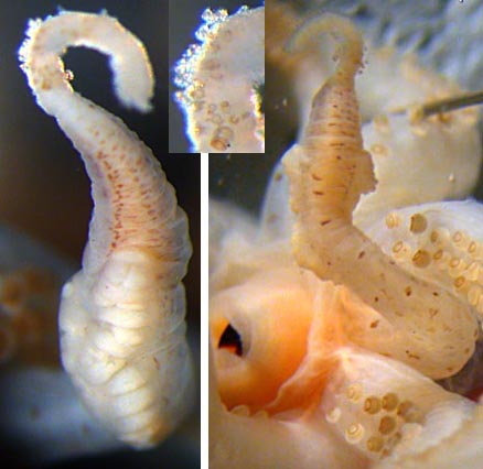
Figure. Tentacles of M. atlantica, holotype. Left - Oral view of the glandular region of the left tentacle. Note the broken tip where filament attaches. A full view of this tentacle is seen in the oral view of the arms above. Middle - Oral-oblique view of the glandular region of the right tentacle and a portion of the distal region (broken at end) bearing small suckers. Insert - Distal-tentacle showing suckers more clearly. Right - Aboral view of the right tentacle. Photographs from Vecchione and Young (2006), modified.

Figure. Side view of the head of M. atlantica showing the eyelid and olfactory organ. Photograph by R. Young.
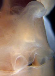
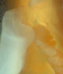
Figure. Funnel/mantle locking-appraus of M. atlantica, holotype. Left - Frontal-oblique view of the funnel component. Right - Side view of the mantle component. Photographs by R. Young.
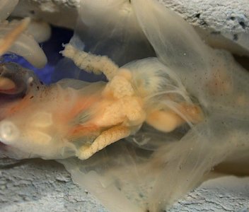
Figure. Ventral view of the mantle cavity of M. atlantica, holotype. Photograph by R. Young.


