


Cardiapoda richardi
Roger R. Seapy

Introduction
Cardiapoda richardi is distinguished from C. placenta by the following features: a smaller adult body size (to about 30 mm); eight gills located at the entrance to the mantle cavity; lens of the eye rests in a depression in a black base of hemispherical shape; fin sucker present only in males; low dorsal crest on the tail; retractable brown, membranous structure on the mid-ventral surface of the tail; reddish-brown, filamentous tail extension; and, radula with the central rachidian tooth bearing three pointed cusps, of which the outer ones are about one-third shorter than the middle one.
Brief Diagnosis
A species of Cardiapoda with:
- Eight gills located at the entrance to the mantle cavity
- Lens of eye rests in a depression in a solid black base of hemispherical shape
- Fin sucker only in males
- Tail with a low dorsal crest
- Ventral surface of tail with a retractable, brown membranous structure
- Tail terminates in a reddish-brown, filamentous extension
- Central rachidian teeth each bear three pointed cusps, of which the outer ones are about one-third shorter than the middle one
Characteristics
- Body morphology
- Eight gills located at entrance to mantle cavity
- Tentacles elongate and of equal length
Click on an image to view larger version & data in a new window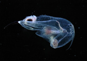
Figure. Frontal view of Cardiapoda richardi. © L. Madin
- Numerous, small white spots cover the body. In side profile the spots appear elevated, like very low tubercles. Each spot appears to consist of a cluster of minute, white specks
Click on an image to view larger version & data in a new window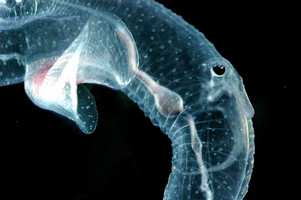
Figure. Right side of body in Cardiapoda richardi. © L. Madin
- Lens of eye rests in a cup-like depression in a large, pigmented base
Click on an image to view larger version & data in a new window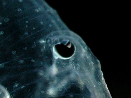
Figure. View of right side of head region and eye in Cardiapoda richardi. © L. Madin
- Tail with a low dorsal crest (a brown membranous structure or expansion that can be retracted into an elongate mid-ventral groove) and a terminal reddish-brown, filamentous extension that is highly contractile (compare the length of the tail extension in the two title illustrations). Thiriot-Quiévreux (1975) reported the presence of the membranous structure on the tail in larvae of the species
Click on an image to view larger version & data in a new window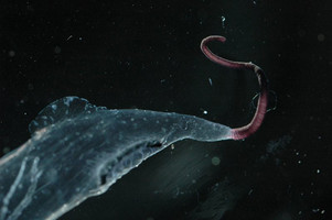
Figure. Ventro-lateral view of tail in Cardiapoda richardi. © L. Madin
- Radula
- The radula of a 9-mm juvenile Cardiapoda richardi was illustrated by Thiriot-Quiévreux (1975; see below). The central rachidian teeth each consist of a middle region with three cusps that are close together at the base; the middle one is of moderate length and has a broad base, the left one is about one-half the length and basal width of the median one, and the right one is very short Click on an image to view larger version & data in a new window
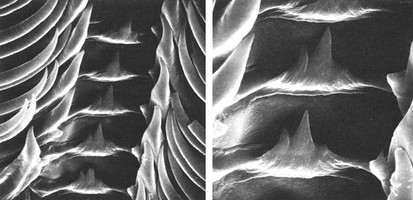
Figure. Scanning electron micrographs of the radula from a juvenile Cardiapoda richardi at low (left) and high (right) magnfications. Photographs from Thiriot-Quiévreux (1975, Fig. 5D,E). © 1975 C. Thiriot
- Adult radular morphology is not recorded in the literature. The photograph below (provided by Orso Angulo, Universidad Autónoma de Baja California Sur) is from a 20-mm individual. In comparison with the central rachidian teeth in the juvenile radula described above, the middle cusp remains the longest of the three cusps, but both of the outer ones are about one-third shorter and of comparable length. In contrast, the three cusps of the central rachidian teeth in C. placenta are of comparable length, are spaced further apart from each other, and the outer cusps flair somewhat outward (see the C. placenta page) Click on an image to view larger version & data in a new window
Figure. High-magnification photograph of a section of the radula from a 20-mm adult Cardiapoda richardi. © Orso Angulo
- The radula of a 9-mm juvenile Cardiapoda richardi was illustrated by Thiriot-Quiévreux (1975; see below). The central rachidian teeth each consist of a middle region with three cusps that are close together at the base; the middle one is of moderate length and has a broad base, the left one is about one-half the length and basal width of the median one, and the right one is very short
- Shell
- Adult shell not characterized in the literature. Spoel (1976:158) stated "A shell is never described for this species, but a coil in the liver top of small specimens proves that a small shell must be present also after the veliger stage"
- The appearance of the early post-metamorphic shell was observed by Orso Angulo (Universidad Autónoma de Baja California Sur; unpublished). The photograph below is of an early juvenile specimen with its visceral nucleus and digestive gland covered by the transparent shell, which has a short, raised keel
Click on an image to view larger version & data in a new window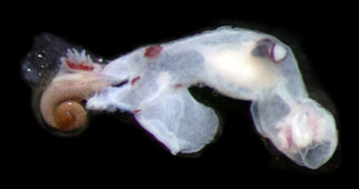
Figure. Early post-metamorphic specimen of Cardiapoda richardi (body length = 5.7 mm). © 2008 Orso Angulo
- From the above specimen, Angulo removed the shell and obtained the scanning electron micrograph below. In marked contrast with the shell of Cardiapoda placenta, in which the teleoconch is produced at right angles to the larval shell aperture (see the C. placenta page), the teleoconch grows directly outward (in the same plane) from the larval shell aperture; similar to Carinaria. Also, the teleoconch in C. placenta lacks a keel, while in C. richardi a keel is present that is produced at the beginning of the teleoconch. The presence of a keel that develops at the beginning of the teleoconch is strikingly similar to what is seen in Carinaria (see the color photographs of the visceral nucleus and shell on the C. galea and C. lamarcki pages)Click on an image to view larger version & data in a new window
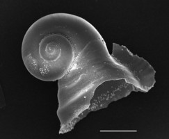
Figure. Shell from a 5.7-mm Cardiapoda richardi, viewed from the right side. Scale bar = 0.5 mm. © 2008 Orso Angulo
- Larva
- Larval pigmentation was described from specimens collected in the western North Atlantic by Thiriot-Quiévreux (1975) and the North Pacific (Hawaiian waters) by Seapy and Thiriot-Quiévreux (1994). In the former study the body was characterized as having little or no pigmentation; digestive gland yellow; velar lobes with a thin brown border; right tentacle brown, left one colorless. Larvae observed from Hawaiian waters differed by having a light yellow body coloration, light brown digestive gland, and velar lobes lacking terminal pigmentation
- Larval shell globular, consisting of about 3-1/4 whorls (see first image below). Spire whorls smooth except for a pair of elevated spiral ridges on the second whorl (first image below); in agreement with scanning electron micrographs of the shell spire in Thiriot-Quiévreux (1975, figs. 1K,L). Aperture wide, slightly greater than 1/2 shell diameter (second image below). Umbilicus open, with about 13 elevated, radiating striae on wall (third image below)
Click on an image to view larger version & data in a new window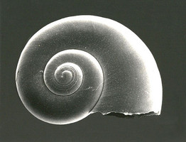
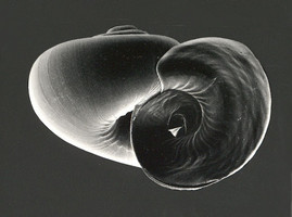
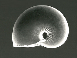
Figure. Scanning electron micrographs of the larval shell of Cardiapoda richardi, viewed from the right side, aperture, and left side (left, center and right, respectively. Shell diameter = 0.6 mm. © R. R. Seapy
- Larval pigmentation was described from specimens collected in the western North Atlantic by Thiriot-Quiévreux (1975) and the North Pacific (Hawaiian waters) by Seapy and Thiriot-Quiévreux (1994). In the former study the body was characterized as having little or no pigmentation; digestive gland yellow; velar lobes with a thin brown border; right tentacle brown, left one colorless. Larvae observed from Hawaiian waters differed by having a light yellow body coloration, light brown digestive gland, and velar lobes lacking terminal pigmentation
References
Lalli, C. M. and R. W. Gilmer. 1989. Pelagic snails. The biology of holoplanktonic gastropod snails. Stanford: Stanford University Press. 259 pp.
Richter, G. and R. R. Seapy. 1999. Heteropoda, pp. 621-647. In: D. Boltovskoy (ed.), South Atlantic Zooplankton. Leiden: Backhuys Publishers.
Seapy, R. R. and C. Thiriot-Quiévreux. 1994. Veliger larvae of Carinariidae (Mollusca: Heteropoda) from Hawaiian waters. Veliger 37: 336-343.
Tesch, J. J. 1949. Heteropoda. Dana Report 34, 54 pp., 5 plates.
Thiriot-Quiévreux, C. 1975. Observations sur les larves et les adultes de Carinariidae (Mollusca: Heteropoda) de l'Océan Atlantique Nord. Marine Biology 32: 379-388.
Title Illustrations

| Scientific Name | Cardiapoda richardi |
|---|---|
| Location | Florida Current |
| Specimen Condition | Live Specimen |
| Sex | Female |
| Life Cycle Stage | adult |
| View | left side |
| Size | 65 mm |
| Copyright | © Ronald Gilmer |
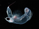
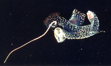
| Scientific Name | Cardiapoda richardi |
|---|---|
| Location | Sargasso Sea |
| Specimen Condition | Live Specimen |
| Sex | Female |
| Life Cycle Stage | adult |
| View | ventral |
| Copyright | © L. Madin |
About This Page

California State University, Fullerton, California, USA
Correspondence regarding this page should be directed to Roger R. Seapy at
Page copyright © 2008
 Page: Tree of Life
Cardiapoda richardi .
Authored by
Roger R. Seapy.
The TEXT of this page is licensed under the
Creative Commons Attribution License - Version 3.0. Note that images and other media
featured on this page are each governed by their own license, and they may or may not be available
for reuse. Click on an image or a media link to access the media data window, which provides the
relevant licensing information. For the general terms and conditions of ToL material reuse and
redistribution, please see the Tree of Life Copyright
Policies.
Page: Tree of Life
Cardiapoda richardi .
Authored by
Roger R. Seapy.
The TEXT of this page is licensed under the
Creative Commons Attribution License - Version 3.0. Note that images and other media
featured on this page are each governed by their own license, and they may or may not be available
for reuse. Click on an image or a media link to access the media data window, which provides the
relevant licensing information. For the general terms and conditions of ToL material reuse and
redistribution, please see the Tree of Life Copyright
Policies.
- First online 12 February 2008
- Content changed 12 September 2008
Citing this page:
Seapy, Roger R. 2008. Cardiapoda richardi . Version 12 September 2008. http://tolweb.org/Cardiapoda_richardi/28745/2008.09.12 in The Tree of Life Web Project, http://tolweb.org/


