LOWER BEAK
We examined beaks of M. psychrophila from two individuals (99, 100 mm ML). The following characters are taken from Clarke (1986). Means are used, when possible, for measurements.
- Pigmentation - Slightly fainter than normal.
- Pigmentation pattern - Fully pigmented at 99 mm ML.
- (Wing length)/(visible rostral edge) (profile) - 2.43
- Height/baseline (profile) - 0.90
- (Length from rostral tip to wing tip projected on baseline)/baseline (profile) - 0.25
- (Hood length)/(crest length) (profile) - 0.53
- (Crest length)/baseline (profile) - 0.56
- (Rostral base)/(visible rostral edge) (profile) - 0.89
- (Hood length)/(visible rostral edge) (profile) - 0.96
- Rostral width ratio [LRL/(length between jaw angles)] - 1.27
- Wing angle (= visible jaw angle in profile) - Obtuse.
- Shape of rostral edge - Nearly straight but with tiny hook.
- Rostrum (profile) - Broad taper.
- Hood distance from crest - Normal.
- Crest - Straight or slightly curved.
- Lateral wall notch - Slight.
- Hood-wing - Broad.
- Hood notch - Moderate.
- Lateral walls to free corner - Well separated with increasing but low divergence from one another.
- Hood midline - Very slightly curved.
- Hood surface without prominent longitudinal ridges or grooves.
- Wing fold - Low, rounded, barely obscures jaw angle in profile.
- Shoulder - Shoulder blades shape in advanced stage, edges barely project.
- Shoulder groove - Virtually absent.
- Jaw angle - Nearly right angle or obtuse, without associated step or transparent strip.
- Jaw edge extension without step and with prominent angle point extending well past shoulder padding; jaw edge extension without clear strip between it and shoulder blade.
- Jaw edge in cross-section - Sharp.
- Crest in cross-section - Narrow.
- Lateral wall - Distinct lateral-wall fold capped by solid ridge that usually overhangs base anteriorly, ridge nearly reaches posterior margin of lateral wall; lateral wall fold, with anterior sharp, medial groove and steeply sloping sides, extends to posterior margin of lateral wall.

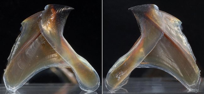
Figure. Left and right side views of the same lower beak of M. psychrophila 100 mm ML, 3.0 mm LRL, immature female, South Atlantic: 53°S, 38°W. Photographs by R. Young.
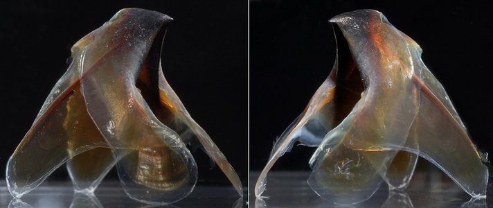
Figure. Left and right side-oblique views of the same lower beak of M. psychrophila 100 mm ML, immature female. Photographs by R. Young.
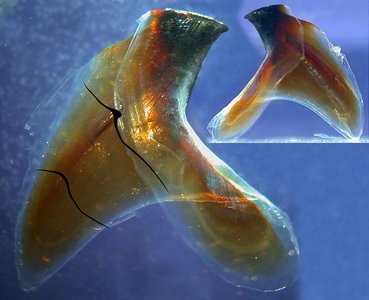
Figure. Side view of the lower beak of M. psychrophila 99 mm ML, 3.0 mm LRL, Antarctic waters at 58°S, 77°W. The beak has been cut in half and laid flat with drawn cross-sections of the lateral wall overlayed where the cuts occurred. Inset - Same beak, intact, upright side view. Photographs by R. Young.
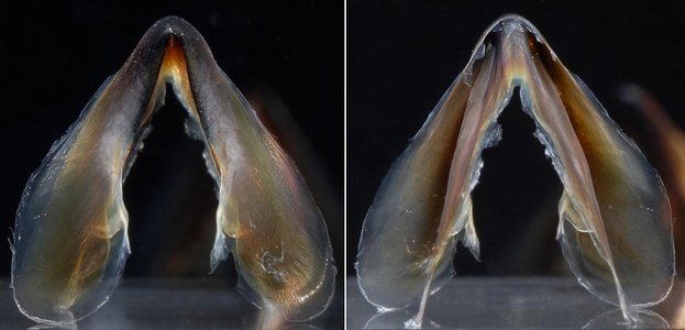
Figure.Front (left) and rear (right) views of the same lower beak of M. psychrophila 100 mm ML, immature female. Photographs by R. Young.
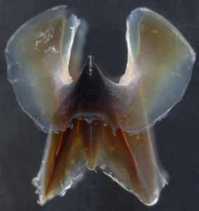
Figure. Top view of the lower beak of M. psychrophila 100 mm ML, immature female. Photographs by R. Young.
Comments
To be added.
UPPER BEAK
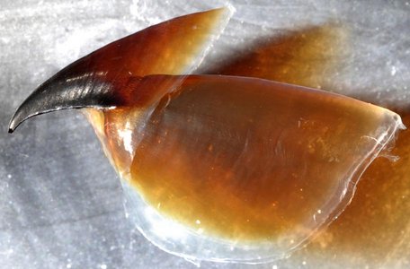
Figure. Side view of the upper beak of M. psychrophila 100 mm ML, immature female. Photographs by R. Young.
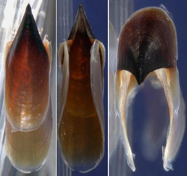
Figure. Various views views of the same upper beak of M. psychrophila 100 mm ML, immature female. Left - Top view. Middle - Bottom view. Right - Front view. Photographs by R. Young.
Comments
To be added.


