- Arms: Arm IV sucker
 Click on an image to view larger version & data in a new window
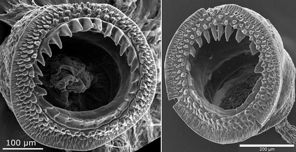
Figure. Oral view of arm IV suckers of M. dentata. Left - Sucker 21, 48 mm ML. Right - Sucker taken at 70% of the arm length from the arm base, 135 mm ML. Photograph by R. Young.
- Tentacles: Club-base suckers Click on an image to view larger version & data in a new window
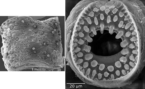
Figure. Left - Oral view of club of M. dentata, 135 mm ML, near club base showing low-density arrangement of suckers. Right - High magnification of one sucker from this region of the club.
- Tentacles: Mid-club suckers
Click on an image to view larger version & data in a new window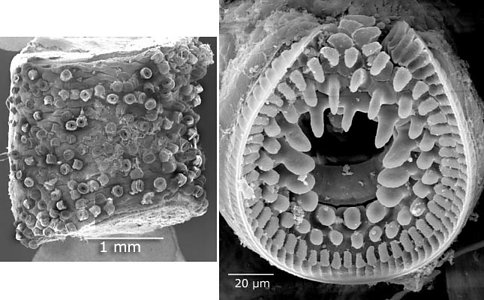
Figure. Left - oral view of club in the midregion of the club of M. dentata, 135 mm ML, showing increased sucker density. Right - High magnification of one sucker from this region of the same club showing the larger sucker size and larger size of the inner lateral knobs. Photographs by R. Young.
- Tentacles: Distal club suckers
Click on an image to view larger version & data in a new window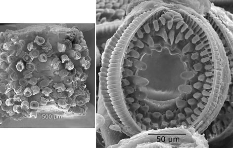
Figure. Left - oral view of club in the distal region of the club of M. dentata, 135 mm ML, showing increased sucker density. Right - High magnification of one sucker from this region of the same club showing the larger sucker size and larger size of the inner lateral knobs. Also the sucker is slightly more elongate than in the basal-club sucker. Photographs by R. Young.
- Tentacles: Mid-club suckers at the side
Click on an image to view larger version & data in a new window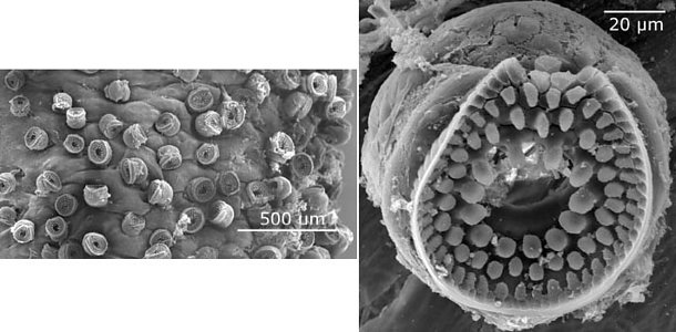
Figure. Left - Side view of the mid-club region of M. dentata. The left side of the image approaches the protective membrane. A sucker-size gradation is present across the club from generally small suckers near the club margin to larger suckers toward the midoral region of the club. Right - Oral view of a small, near-marginal sucker which shows similarities in the size of the lateral pegs to the suckers of the proximal section. Photographs by R. Young. Photograph by R. Young.
Mastigoteuthis dentata: Scanning electron micrographs of suckers
Richard E. YoungAbout This Page

University of Hawaii, Honolulu, HI, USA
Correspondence regarding this page should be directed to Richard E. Young at
Page copyright © 2006
 Page: Tree of Life
Mastigoteuthis dentata: Scanning electron micrographs of suckers
Authored by
Richard E. Young.
The TEXT of this page is licensed under the
Creative Commons Attribution-NonCommercial License - Version 3.0. Note that images and other media
featured on this page are each governed by their own license, and they may or may not be available
for reuse. Click on an image or a media link to access the media data window, which provides the
relevant licensing information. For the general terms and conditions of ToL material reuse and
redistribution, please see the Tree of Life Copyright
Policies.
Page: Tree of Life
Mastigoteuthis dentata: Scanning electron micrographs of suckers
Authored by
Richard E. Young.
The TEXT of this page is licensed under the
Creative Commons Attribution-NonCommercial License - Version 3.0. Note that images and other media
featured on this page are each governed by their own license, and they may or may not be available
for reuse. Click on an image or a media link to access the media data window, which provides the
relevant licensing information. For the general terms and conditions of ToL material reuse and
redistribution, please see the Tree of Life Copyright
Policies.


