| 1 | Abdomen linear, with segments VIII-IX not modified; segment IX with pair of tubular urogomphi (rarely reduced or absent) | most Carabidae |
| - | Abdomen upcurved, with segments VIII-IX (mostly epipleurites and tergites) highly modified, transformed into a transverse sclerotized terminal disk composed of flat plates; segment IX with pair of branched or plate-like urogomphi (Paussinae)  Click on an image to view larger version & data in a new window 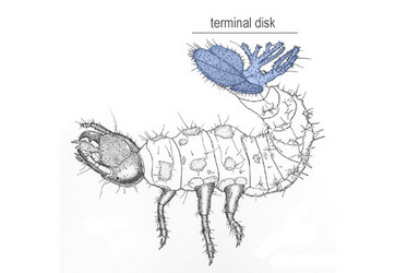 Terminal disk. | 2  |
 2 2 | Head hyperprognathous (bent upwards); neck constricted; coronal suture present; sensorial appendage small and conical on antennomere III; mandibles long and slender, sickle-shaped; urogomphi multi-lobed; tarsi with 2 claws; pygidium positioned between urogomphi near the middle of the terminal disk (Metriini, Ozaenini) | 3 |
| - | Head prognathous; neck not constricted; coronal suture absent; sensorial appendage large, bulbous or conical on antennomere III; mandibles short and broad, subtriangular; urogomphi flattened, plate-like, not lobate; tarsi with 1 claw; pygidium positioned ventral to urogomphi and terminal disk (Paussini) Click on an image to view larger version & data in a new window 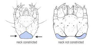 Neck shape. 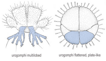 Urogomphi shape.  Position of pygidium. | 8 |
| 3 | Anterior margin of frontoclypeolabrale with four small subequal teeth, positioned transversely just behind the anterior margin of frontal sclerite; stipes of maxillae without a basal tooth; urogomphi with 5 lobes, lobe A short, lobe D placed at the common base of lobes B-C, lobe E undivided | Metrius |
| - | Anterior margin of frontoclypeolabrale not toothed, smooth transverse keel present anteriorly on frontal sclerite; stipes of maxillae with an acute basal tooth at inner side; urogomphi with 6 or more lobes, lobe A elongate, lobe D at the base of lobe E; lobe E subapically divided into E1 and E2 lobes (Ozaenini) Click on an image to view larger version & data in a new window 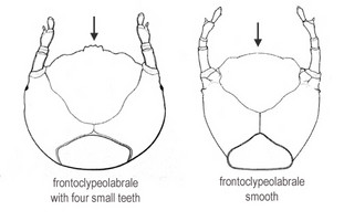 Frontoclypeolabrale shape. 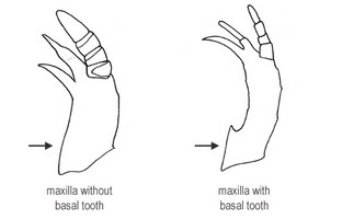 Maxilla shape. 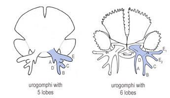 Number of urogomphal lobes. | 4 |
| 4 | Transverse keel just behind anterior margin of frontoclypeolabrale; labral tooth small, anteriorly truncate or partially bilobate, partially covered by the transverse keel in dorsal view; prementum subrectangular or subquadrate, with parallel sides; urogomphi antler-like with long and slender lobes, lobes D and E1 rounded at tip; dorsal plates of terminal disk flat or only slightly convex without protruding sensorial areas | 5 |
| - | Transverse keel well behind anterior margin of frontoclypeolabrale; labral tooth wide, slightly rounded anteriorly, clearly visible in dorsal view; prementum tapered from the base to the apex; urogomphi with short, flattened and partially fused lobes, lobes D and E1 pointed at tip; dorsal plates of terminal disk concave with large, protruding sensorial areas near the middle Click on an image to view larger version & data in a new window 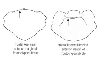 Position of transverse keel. 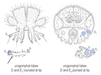 Urogomphal lobes D and E. 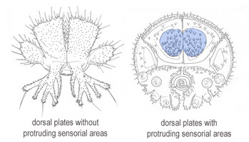 Surface of dorsal plate. | Physea |
| 5 | Prementum subrectangular, longer than wide; ligula dome-like, moderately protruding; antennomere IV short and cylindrical; second galeomere conical | Pachyteles |
| - | Prementum subquadrate or slightly transverse; ligula elongate; antennomere IV long and clavate; second galeomere stick-like, elongate and cylindrical Click on an image to view larger version & data in a new window 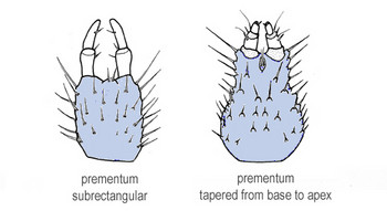 Prementum shape. 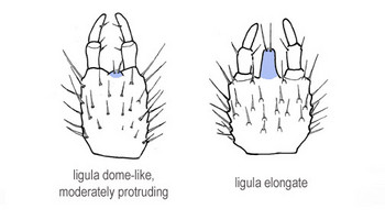 Ligula length. | 6 |
| 6 | Urogomphi with lobes A and E2 flattened, truncate and partially divided at apex (according to the drawings of Paulian, 1947); lobe B undivided | Sphaerostylus (Afrozaena) |
| - | Urogomphi with lobes A and E2 cylindrical, tapering to the tip and undivided; lobe B divided into B1 and B2 lobes Click on an image to view larger version & data in a new window 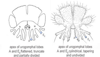 Urogomphal lobes A and E. 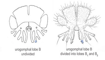 Urogomphal lobe B. | 7 |
| 7 | Lacinia slightly longer than galea; dorsal plates of terminal disk subtrapezoidal, distinctly wider at apex than base, medially adjacent; lateral plates wide and short; lobe A slender, about as long as lobe B; lobe D slender and long | Itamus |
| - | Lacinia shorter than galea; dorsal plates of terminal disk subrectangular, only slightly wider at apex than base, deeply divided medially; lateral plates elongate and slender; lobe A robust, distinctly shorter than lobe B; lobe D very small Click on an image to view larger version & data in a new window 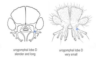 Urogomphal lobe D length. 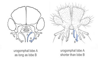 Urogomphal lobe A length. | Goniotropis |
| 8 | Several setae on dorsal surface of the head, thoracic tergites and of perimeter of terminal disk clavate and riddled (sensilla S-VIII); lacinia present, apically with strong hook-like spines along occlusal edge; antennomere IV with stick-like, apical sensory appendix; mandible without prostheca; leg with articulated trochanter, femur, tibia and tarsus; urogomphi with irregularly raised margin | Arthropterus |
| - | Setae on dorsal surface of the head, thoracic tergites and perimeter of terminal disk not clavate; lacinia absent; antennomere IV simple, without stick-like, apical sensory appendix; mandible with digitiform prostheca; leg with fused trochanter, femur, tibia and tarsus; urogomphi with regularly rounded margin Click on an image to view larger version & data in a new window 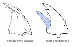 Mandible shape. 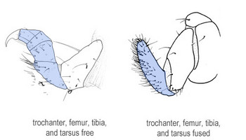 Fusion of leg articles. | 9 |
| 9 | First maxillary palpomere basally articulated with stipes, resulting in a 4-jointed palp; sutures between trochanter, femur, tibia and tarsus visible; sensilla S-I with inflated apex; sensilla S-II on margin of terminal disk absent | Platyrhopalopsis |
| - | First maxillary palpomere basally fused with stipes, resulting in a 3-jointed palp; trochanter, femur, tibia and tarsus completely fused, without sutures; sensilla S-I with simple truncate apex; sensilla S-II on margin of terminal disk present Click on an image to view larger version & data in a new window 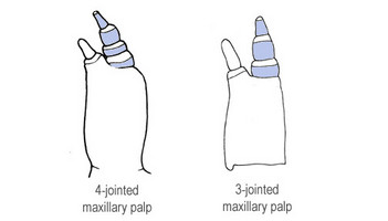 Number of maxillary palp articles. 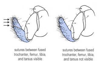 Sutures of fused leg articies. | Paussus |
Illustrated Key to Paussinae Larvae
Wendy Moore and Andrea Di GiulioReferences
Moore, W. and A. Di Giulio. 2006. Description and behaviour of Goniotropis kuntzeni larvae (Coleoptera: Carabidae: Paussinae: Ozaenini) and a genus-level key to Paussinae larvae. Zootaxa 1111:1-19.
About This Page
Drawings © Andrea Di Giulio
Wendy Moore

University of Arizona, Tucson, Arizona, USA
Università degli Studi "Roma Tre", Roma, Italy
Correspondence regarding this page should be directed to Wendy Moore at
Page copyright © 2007 Wendy Moore and
 Page: Tree of Life
Illustrated Key to Paussinae Larvae
Authored by
Wendy Moore and Andrea Di Giulio.
The TEXT of this page is licensed under the
Creative Commons Attribution-NonCommercial-ShareAlike License - Version 3.0. Note that images and other media
featured on this page are each governed by their own license, and they may or may not be available
for reuse. Click on an image or a media link to access the media data window, which provides the
relevant licensing information. For the general terms and conditions of ToL material reuse and
redistribution, please see the Tree of Life Copyright
Policies.
Page: Tree of Life
Illustrated Key to Paussinae Larvae
Authored by
Wendy Moore and Andrea Di Giulio.
The TEXT of this page is licensed under the
Creative Commons Attribution-NonCommercial-ShareAlike License - Version 3.0. Note that images and other media
featured on this page are each governed by their own license, and they may or may not be available
for reuse. Click on an image or a media link to access the media data window, which provides the
relevant licensing information. For the general terms and conditions of ToL material reuse and
redistribution, please see the Tree of Life Copyright
Policies.


