

Figure. Dorsal view of A. mangoldae, paratype (SBMNH 369535, 100 mm ML, mature male), preserved. Photograph by R. Young.
- Arms
- Largest arm suckers with ca. 9 truncated teeth on distal half of ring.
Click on an image to view larger version & data in a new window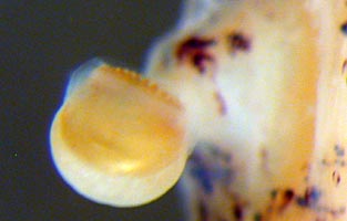
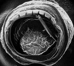
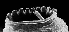
Figure. Arm suckers of A. mangoldae. Top left - Distal view of a large arm III sucker, 100 mm ML, mature male, preserved. Photograph by R. Young. Top right - Oral view of one arm sucker (sucker 10, arm II, holotype), scanning electron micrograph (SEM). A piece of debris lies inside the sucker acetabulum. Bottom - Oblique view of the same arm II sucker, SEM. Note that the teeth project vertically outward from the face of the sucker. Photographs by R. Young.
- Arms IV longest but relative size decreases with increasing squid size.
- Arm IV suckers much smaller than arm III suckers.
Click on an image to view larger version & data in a new window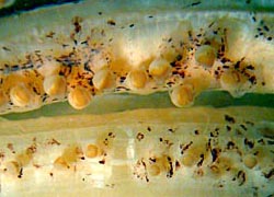
Figure. Oral views of arms III (top) and IV (bottom) of A. mangoldae at the same distance from the arm bases, 100 mm ML, mature male, preserved. Photograph by R. Young.
- Sucker stalks with broad bases
- Tentacles
- Suckers with ca. 8 large, truncated teeth on distal half of inner ring grading to ca. 17 small, truncated teeth on proximal half.
Click on an image to view larger version & data in a new window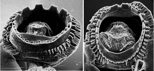
Figure. Oral views from different angles of two large club suckers from A. mangoldae, holotype, scanning electron micrographs. Note that the teeth project vertically outward from the face of the sucker. Photographs by R. Young.
- The club sucker stalks consists of a single portion.
- Stalks from the medial and lateral series approxmately the same length.
Click on an image to view larger version & data in a new window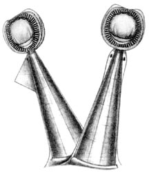
Figure. Oral views of club suckers and stalks of A. mangoldae. Left - A pair of club suckers with stalks from the medial and lateral sucker series, NMNH 729749, holotype. Flag on lateral stalk was not always present. Drawing by A. Hart. Right - Most of the distal portion of the club, holotype, preserved. Photograph by R. Young.
- Club with 13 trabeculae in each distal protective membrane; 24-25 trabeculae in each proximal protective membrane.
- Head
- Head elongate with long neck and brachial pillar.
- Olfactory organ on long stalks and postioned well posterior to eyes at level of posterior end of cephalic cartilage.
- Radula
- Radula with strong secondary cusps on both the rhachidian and first lateral teeth.
- Beaks
- Upper beak with strongly recessed shoulder with sharp ridge on oral and aboral sides of shoulder. Oral ridge extends well onto pallet. Ridges absent from aboral surface of pallet. Rostrum short and curved at tip.
- Lower beak with wing fold obscuring beak angle and extending nearly to tip of rostrum. Lateral wall with weak oblique fold.
- Funnel
- Funnel valve present. Click on an image to view larger version & data in a new window
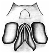
Figure. Ventral view of and funnel organ of A. mangoldae, NMNH 729752, with the funnel wall cut and spread open. Drawing by A. Hart.
- Mantle
- Mantle tissue with gelatinous consistency and filled with small vesicles.
- Mantle musculature terminates in anterior third of fins.
- Fins
- Combined fin width exceeds fin length.
- Photophores
- Terminal aboral club photophore small.
- Small embedded photophores on aboral surface of club laterally near protective membranes. About 8 in distal section of club on each side and 5 in proximal section on each side.
Click on an image to view larger version & data in a new window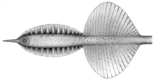
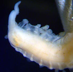
Figure. Aboral view of the club of A. mangoldae. Top - NMNH 729749, holotype. Drawing by A. Hart. Bottom - Distal portion of club, holotype, preserved. Photograph by R. Young.
- Eye photophore
- Visceral photophores
- Visceral photophores absent.
- Pigmentation
- Pigmentation in typical chromatophores; chromatophores scattered on body and buccal membrane.
- Viscera
- Penis long and slender in mature male.
Click on an image to view larger version & data in a new window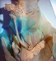
Figure. Ventral view of the mantle cavity and funnel of A. mangoldae, paratype, 100 mm ML, mature male, preserved. Note the long, slender penis extending from the gill base past the blue-stained funnel locking-apparatus and carrying a lobate tip.
- Measurements
Holotype NMNH 729749 Clarke 70-12-31 Clarke 70-12-31 Clarke 70-12-31 Paratype SBMNH 369535
Sex Imm. male -- -- -- Mature male
Mantle length 80 128 120 82 100 Fin length 30 56 55 34 53 Fin width 37 72 66 39 67 Eye diameter Damaged Damaged
Damaged
Damaged
ca. 17
Arm I, length 33 76 69 44 65 Arm II, length 41 86 73 46 75 Arm III, length 40 85 79 48 72 Arm IV, length 70 100 90+ 95 86 Club length 15 Missing Missing Missing Missing - Additional measurements of the holotype of A. mangoldae: Mantle width - 13 mm, Head length and width - approximately 30 and 9 mm but damaged; Number of arm suckers of proximal 50% of arms III and IV - 25, 24; Sucker size of largest suckers of arms I-IV respectively - 0.6, 0.6, 0.7, 0.5 mm.
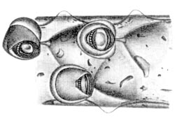
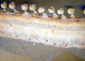
Figure. Arm III suckers of A. mangoldae. Left - Oral view of three suckers and their stalks, NMNH 729752. Drawing by A. Hart. Right - Dorsal view of mid-arm suckers, 100 mm ML, mature male, preserved. Photograph by R. Young.
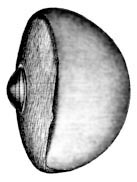
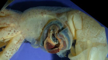
Figure. The eyeball of A. mangoldae. Left - Ventral view, USNM 729752, showing large photophore. Drawing by A. Hart. Right - Side view of a damaged head and eye. The iridescent tissue has pulled free of the eyeball and the lower portion, appearing dark red, is the photophore that was originally positioned on the ventral surface of the eyeball, paratype, 100 mm ML, mature male, preserved. Photograph by R. Young.
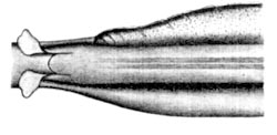
Figure. Ventral view of the intestine of A. mangoldae, NMNH 729752. Above the intestine, in the drawing, is the ink sac and below the intestine, in the drawing, is the cephalic vein.


