- Mantle cavity
- The arrow points to the lateral mantle opening. The mantle cavity is colored blue. The funnel is very large and is entirely fused ventrally to the mantle. The digestive gland attaches ventrally to the mantle via the mantle adductor muscles.
 Click on an image to view larger version & data in a new window
Figure. Cut away lateral view of A. pelagicus showing the mantle cavity. Drawing by R. Young.
- Digestive system
- The intestine does not pass over the ventral surface of the digestive gland but passes around the right side of the gland near the ventral surface. Note the dorsal position of the stomach and caecum and the loop in the esophagus that allows the dorsal end of the digestive gland to tilt as the animal rotates; the ventral end is fixed by the mantle adductor muscles.
Click on an image to view larger version & data in a new window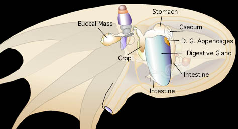
Figure. Cut-away lateral view of A. pelagicus showing the digestive system. Drawing by R. Young.
A. pelagicus has demonstrated in a shipboard aquarium the ability to rotate its eyes and digestive gland to keep them in a vertical orientation. In the photographs below the black line passes through the axis of the gill and provides a reference point. One white lines pass through the axis of the digestive gland and the other through the eye-axis. The photographs show the gill-axis in two different orientations. The orientation of the digestive gland and eye axes (white lines) differ greatly in the two pictures relative to the orientation of the black line (gill axis).
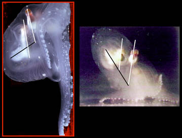
Figure. Left - Side view of A. pelagicus climbing the side of a small shipboard tank. Right - Anterolateral view of the same A. pelagicus sitting on the bottom of the tank. Photographs by R. Young.


