Two hooks (V5 and D5) of Notonykia africanae were examined with the scanning electron microscope (SEM). The relative sizes of the hooks are seen in the photograph below. The largest of all the club hooks was the fifth hook from the proximal end of the club in the ventral series (ie, hook V5, below right) and its partner from the dorsal series is hook D5 (below left). The hooks were not well fixed and were difficult to clean for SEM; hook V5 cracked in the process.

Figure. Frontal views of hooks D5 (left) and V5 (right) of N. africanae, 40 mm ML, NMNH specimen (WH 380-III-71). Photographs by R. Young.

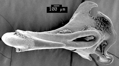
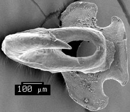
Figure. Top-frontal views of the two hooks (V5 left, D5 right). The grooves of the claw on V5 join and the top portion of the groove wall is absent. A wave-like hair-lip is present on the proximal lip. Both lateral lobes are well developed but one is much larger than the other and both have a strongly hooked-shape. The joining of the claw grooves can barely be seen in the photograph of D5 but is more apparent in the photograph of this hook below. Photographs by R. Young.

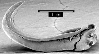
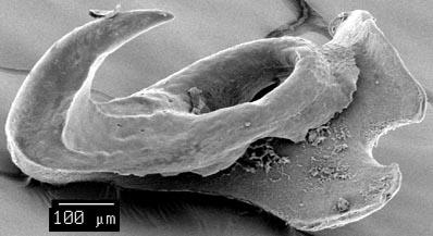
Figure. Proximal-side views of the two hooks (V5 top, D5 bottom). Photographs by R. Young.

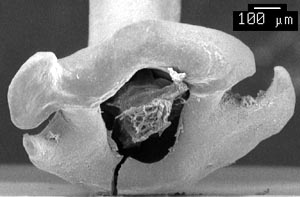
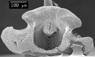
Figure. Attachment-site views of the two hooks (V5 left, D5 right). The bottom orifice of V5 has some tissue that couldn't be removed. Photographs by R. Young.
Comments
The joining of the claw grooves is seen only in this squid and Kondakovia longimana. The hooks of N. africanae are distinct in the hook-shape of the lateral lobes of the ventral hook and the rather slender lateral lobes of the dorsal hook.


 Go to quick links
Go to quick search
Go to navigation for this section of the ToL site
Go to detailed links for the ToL site