


Octopoteuthis deletron
Richard E. Young
Introduction
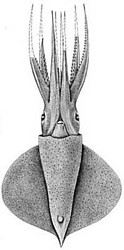
Figure. Ventral view of O. deletron. Drawing from Young (1972).
This species is easily separated from the only other north Pacific species (O. nielseni) by the presence of one rather than two large tail photophores.
Diagnosis
A Octopoteuthis ...
- with a single tail photophore.
- with a temperate Pacific Ocean habitat.
Characteristics
- Arms
- Hooks with accessory cusps that become more prominent with size.
Click on an image to view larger version & data in a new window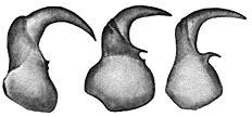
Figure. Side view of large arm III hooks of O. deletron. Left - Ca. 20 mm ML. Middle - 109 mm ML. Right - 167 mm ML. Drawings from Young (1972).
Click on an image to view larger version & data in a new window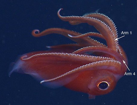
Figure. Side view of a defensive posture of O. deletron. Note all arms have their oral (hook-bearing surfaces) directed forward. In situ photograph taken from an MBARI ROV at 516 m. © 2013 MBARI.
- 3-12 pairs of small suckers near arm tips, each with 6-9 irregular, pointed teeth.
Click on an image to view larger version & data in a new window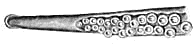

Figure. Left - Oral view of arm III tip of O. deletron, 167 mm ML. Right - Oral view of inner sucker ring from the same arm tip, 5th transverse row from hooks. Drawings from Young (1972).
- Hooks with accessory cusps that become more prominent with size.
- Tentacles
- Absent by 15-25 mm ML.
- Absent by 15-25 mm ML.
- Mantle
- Grooves and ridges (rugosity) on anterior half of mantle present only in maturing or mature females (Hoving, et al., 2012). See photograph at right.
- Grooves and ridges (rugosity) on anterior half of mantle present only in maturing or mature females (Hoving, et al., 2012). See photograph at right.
- Photophores
- Single "tail" photophore.
- Arm organs: series of ca 25 organs near core of each arm III and IV; basal photophore larger and somewhat isolated, on arms II-IV.
- Ventral head organs: one, large, posterior to each eye; elongate, oblique organ near anteromedial margin of each eye.
- Visceral organs: pair near ventral surface of ink sac beneath muscle bands. Click on an image to view larger version & data in a new window
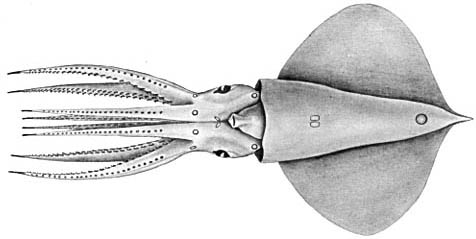
Figure. Ventral view of O. deletron, holotype, 109 mm ML, showing photophore pattern. Drawing from Young (1972).

Figure. Ventrolateral view of the anterior mantle of O. deletron showing female rugosity. In situ photograph taken at a depth of 645m. Photograph © 2013 MBARI.
Comments
Most photophores are embedded in tissue and are difficult to see.Life History
Strong developmental changes occur between ca. 30-40 mm ML that include large changes in eye size, thickness of arms and body, and increases in pigmentation. This is, presumably the transition from the juvenile to the subadult stage. Squid of nearly the same size at this critical stage, therefore, can look like different species. In large specimens, the tail becomes prominent.
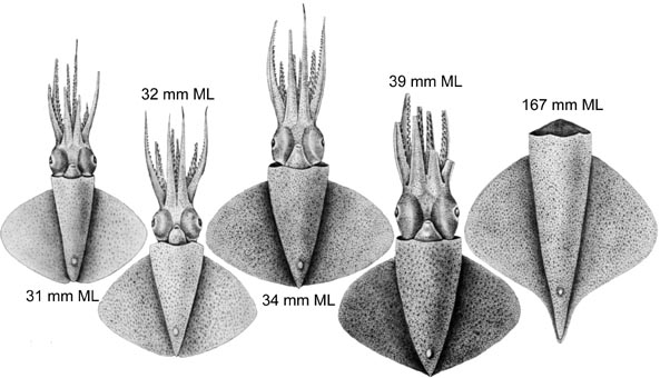
Figure. Ventral views of O. deletron. Left - Four drawings show the marked changes that occur between 30-40 mm ML. Right - The mantle and fins of a large squid showing the distinct tail at this size. Drawings from Young (1972).
Mature males lack a hectocotylus but have a large penis that, apparently, can extend well beyond the mantle opening. Mature eggs are about 2 mm in diameter (Young, 1972). Paralarvae have not been described.
Behavior
Attack autotomy
O. deletron exhibits an unusual defensive behavior known as attack autotomy which was described in detail by S. Bush (2012). The following is extracted from her study. This squid can autotomize an arm leaving a wiggling arm with a flashing photophore in the grasp of a predator. However, for autotomy to occur traction must be involved. That is, only if the arm is securely attached to the predator, will the squid autotomize the arm. This behavior is often part of a sudden attack on the predator followed by a quick retreat. The combination of the behaviors, attack autotomy, is exhibited by only a few other animals, and no other squid (Bush, 2012). The defensive attack behavior, recorded insitu by an MBARI ROV (and part of Bush's study) is seen below:
1. The squid, already on alert by the presence of the ROV, backs into the steel arm of the ROV (arrow). The bottle-brush attached to the arm ROV used to prod squid in various experiments.
2. The squid darts forward with arm-tip photophores lit-up as it splays its arms laterally.
3. The squid abruptly turns back toward its aggressor (the ROV steel arm).
4. With arms still spread and photophores shining, the squid attempts to grasp the steel arm.
5. The attack quickly terminates and the squid darts away.
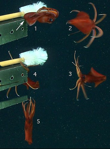
Figure. Single frames from an ROV video taken insitu at a depth of 645 m of O. deletron showing a defensive attack behavior. Photographs 2013 © MBARI.
Flashing photophores
There are two common ways for a squid to control the emission of bioluminescent light: 1) by triggering the photocytes to turn luminesce on or off, or 2) by using a chromatophore shutter over the already luminescing photocytes. O. deletron appears to use the latter for controlling the light emitted by the tips of the arm photophores in some circumstances. This behavior described as "Arm-tip Chromatophores Contracted/Expanded" by Bush et al. (2009) is when the arm-tip photophores flash at variable rates and not together. The function of this behavior is unknown.
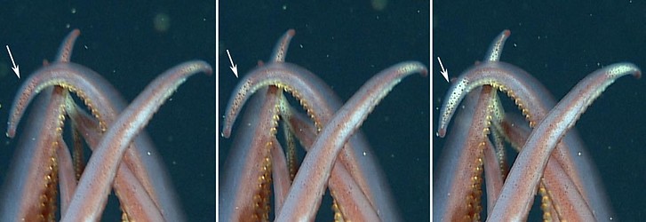
Figure. Control of arm-tip photophores through the use of chromatophore screening in O. deletron. The arrows follow one arm tip from nearly completely covered photophore to a nearly fully exposured luminescing photophore. Figures are from individual video frames taken in situ at 645 m depth by an MBARI ROV. Photographs © 2013 MBARI.
Eyelid blink
Ocotopoteuthids have an unusual modification to the eyelid (A similar eyelid is seen in the related Lepidoteuthis). A posterior-eyelid muscle is extremely large and well defined.
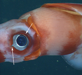
Figure. Ventrolateral view of O. deletron. Same image as seen above (see "3. Mantle") but is re-oriented and cropped. Upper arrow points to the strong, crescent-shaped posterior muscle of the eyelid. Lower arrow points to muscles of the optic sinus what also play a role in eyelid closure. Photograph © 2013 MBARI.

The following video (of a different O. deletron than pictured above) consists of 3 frame-grabs from a longer video to better illustrate how the eyelid closes:
Frame 1 shows the eye wide-open with the lens fully exposed.
Frame 2 shows the posterior eyelid muscle starting to close over the lens.
Frame 3 shows the eyelid nearly closed. Note that the posterior eyelid has moved anteriorly to nearly cover the lens and the optic-sinus muscles have contracted slightly.
Since the basic mechanism of eye closure seen here is similiar to that of some other squids and the time lapse between frame 2 and 3 is not particularity fast (about 1.4 sec.), the reason for the unusually large muscle is not revealed by these photos. However, we have seen, in living Octopoteuthis in ship-board aquaria, the eye close and open so rapidly that the blink could scarcely be detected. If this is the reason for the large posterior eyelid muscle, better documentation is needed.
Distribution
Type locality: Eastern North Pacific at 33°15'N, 118°37'W (off Southern California). This species is found from off Baja California to Alaska, off Northern Peru and possibly off Eastern Honshu (Nesis, 1982/87).
References
Bush, S. L., B. H. Robison and R. L. Caldwell. 2009. Behaving in the dark: Locomotor, chromatic, postural and bioluminescent behaviors of the deep-sea squid Octopoteuthis deletron Young 1972. Biol. Bull. 216: 7-2
Hoving, H. J. T., S. L. Bush and B. H. Robison. 2012. A shot in the dark: same-sex sexual behaviour in a deep-sea squid. Biol. Lett. 8: 287-290.
Nesis, K. N. 1982/87. Abridged key to the cephalopod mollusks of the world's ocean. 385+ii pp. Light and Food Industry Publishing House, Moscow. (In Russian.). Translated into English by B. S. Levitov, ed. by L. A. Burgess (1987), Cephalopods of the world. T. F. H. Publications, Neptune City, NJ, 351pp.
Young, R. E. 1972. The systematics and areal distribution of pelagic cephalopods from the seas off Southern California. Smithson. Contr. Zool., 97: 1-159.
Title Illustrations

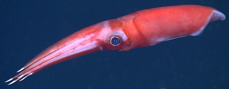
| Scientific Name | Octopoteuthis deletron |
|---|---|
| Location | Northeast Pacific off Northern Oregon at 46.16°N, 124.8°W. |
| Comments | Image courtesy of the Monterey Bay Aquarium Research Institute (MBARI). You must obtain permission from MBARI to use this photo; please contact pressroom@mbari.org for further information |
| Acknowledgements | Susan Von Thun, photo editing, MBARI |
| Specimen Condition | Live Specimen |
| Sex | Female |
| Life Cycle Stage | adult |
| View | Side |
| Copyright | © 2011 MBARI |
About This Page

University of Hawaii, Honolulu, HI, USA
Page copyright © 2016
 Page: Tree of Life
Octopoteuthis deletron .
Authored by
Richard E. Young.
The TEXT of this page is licensed under the
Creative Commons Attribution-NonCommercial License - Version 3.0. Note that images and other media
featured on this page are each governed by their own license, and they may or may not be available
for reuse. Click on an image or a media link to access the media data window, which provides the
relevant licensing information. For the general terms and conditions of ToL material reuse and
redistribution, please see the Tree of Life Copyright
Policies.
Page: Tree of Life
Octopoteuthis deletron .
Authored by
Richard E. Young.
The TEXT of this page is licensed under the
Creative Commons Attribution-NonCommercial License - Version 3.0. Note that images and other media
featured on this page are each governed by their own license, and they may or may not be available
for reuse. Click on an image or a media link to access the media data window, which provides the
relevant licensing information. For the general terms and conditions of ToL material reuse and
redistribution, please see the Tree of Life Copyright
Policies.
- Content changed 21 January 2014
Citing this page:
Young, Richard E. 2014. Octopoteuthis deletron . Version 21 January 2014. http://tolweb.org/Octopoteuthis_deletron/19843/2014.01.21 in The Tree of Life Web Project, http://tolweb.org/


