


Gonatus madokai
Tsunemi Kubodera
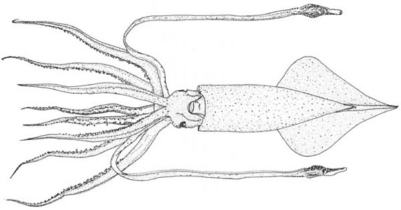
Gonatus madokai: Holotype, ML=329 mm, collected with a salmon gill-net in the sub-surface layer on 6 June, 1977 in the Northwest Pacific Ocean (49-31 N, 168-50 E). Drawing from Kubodera and Okutani (1977).
Introduction
Gonatus madokai was described by Kubodera and Okutani (1977) based on a large holotype (329 mm ML) and two juvenile paratypes (72 mm DML) with three additional specimens (40-63 mm ML) collected from the subarctic waters of the northwestern North Pacific.
Morphological changes with growth of this species were well described (Kubodera and Okutani 1977). G. madokai is one of a few gonatids that can be identified almost throughout its life.
Diagnosis
A Gonatus with ...
- pronouncedly long arms (ca. 90% of ML), long tail (ca. 20% of ML) and soft body.
- large fins (FL>1/2ML), sagittate with round sides (FW=83% of FL).
- tentacle club with a central large hook, a middle-sized hook distal to the central hook and five small hooks in a line proximal to the central hook.
Characteristics
- Arms
- Arms long and thick, but not so muscular.
- Arm III longest (90%ML), arm I shortest (4/5 of arm III).
- Arms III and IV with thin membranous aboral keels along entire lengths.
- Number of hooks and suckers on arm III 46-47 and 52, respectively.
Click on an image to view larger version & data in a new window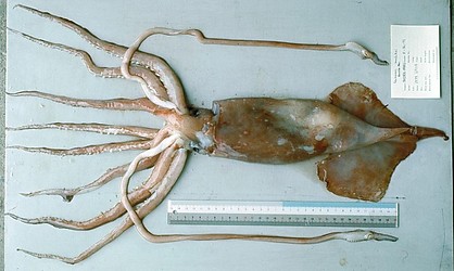
Figure. Ventral view of G. madokai (Holotype), 329 mm ML.
- Tentacles
- Club with 5 small hooks in series proximal to large central hook.
- Club with medium-sized hook distal to large central hook.
- Club suckers of dorsal- and ventral marginal zones merge proximally.
Click on an image to view larger version & data in a new window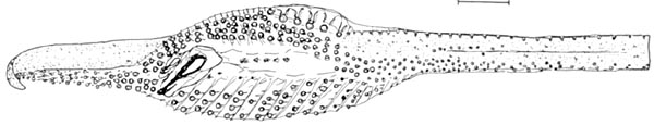
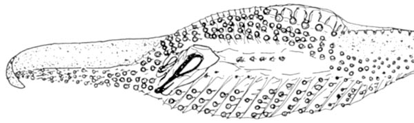
Figure. Oral views of the tentacle and club of G. madokai, 329 mm ML, hologype. Top - Tentacle. Bottom - Enlargement of the tentacular club. Bar 10 mm. Drawings from Kubodera & Okutani (1977)
- Head
- Head almost squarish in shape, slightly narrower than the mantle opening.
- Funnel cartilage lanceolate in shape, nuchal cartilage rectangular with three grooves.
- Dorsal funnel organ inverted V-shaped, ventral ones oval in shape.
- Beaks. Information on the beaks of G. madokai can be found here.
- Radula composed of 5 rows of teeth. Click on an image to view larger version & data in a new window
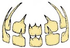
Figure. Radula of G. madokai. Drawing from Kubodera & Okutani (1977).
- Fins and tail
- Fins large sagittate, fin length > 1/2 ML, fin width about 4/5 FL.
- Tail long gelatinous, about 22% of ML.
- Photophores
- Photophores absent.
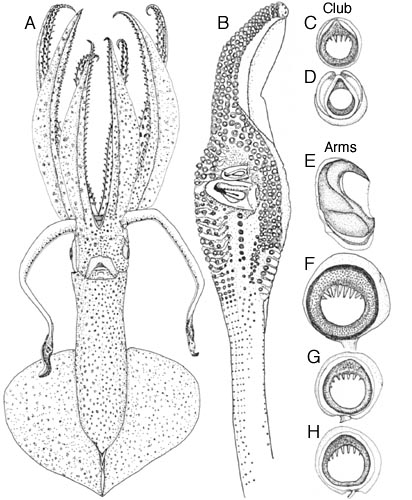
Figure. Juvenile G. madokai (Paratype#1: 72mm ML). A - Ventral view. B - Oral view of right tentacular club. C - Oral view of dactylus sucker. D - Oral view of medial sucker proximal to the central hook. E - Side view of arm III hook. F - Oral view of arm III sucker. G - Oral view of arm IV sucker, medial series. H - Oral view of arm IV sucker, marginal series. Drawings from Kubodera and Okutani (1977).
Life History
Morphological changes with growth of G. madokai were reported by Kubodera and Okutani (1977). Paralarvae and juveniles of G. madokai ranging 8-72mm ML may be identified by having a swollen bell-shaped mantle, trapezoidal head, which is constricted at the base of the arms making eyes protuded in smaller specimens, and slender long arms and tentacles.
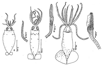
Figure. Morphological changes with growth of Gonatus madokai. A: ventral view of a 11mm ML, B: tentacle of A, C: ventral view of a 22mm ML, D: tentacle of C, E: ventral view of 40mm ML, F: tentacle of E. Drawings from Kubodera and Okutani (1977)
Paralarvae can be identified by the dorsal-head chromatophore pattern which is Type I (three tear-shaped chromatophores on each side.); the mantle has 18 dorsal and 28 ventral chromatophores (Jorgensen, 2006).
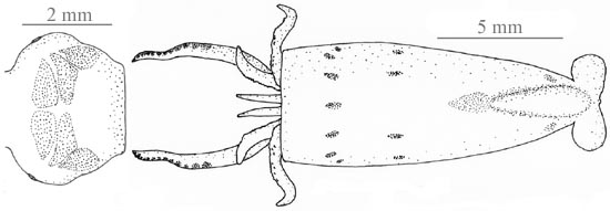
Figure. Dorsal views of the chromatophores of a G. madokai paralarva, 10.9 mm ML, Gulf of Alaska. Left - Head. Right - Paralarva. Drawing from Jorgensen (2007).
Distribution
G. madokai is widely distributed in the subarctic Pacific, and it is especially abundant in the deeper layers of the Okhotsk Sea (Nesis 1997).
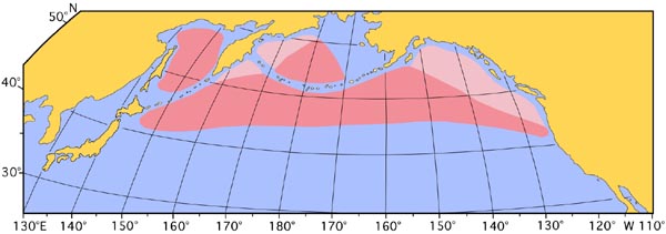
Figure. Distribution of G. madokai based mainly on larval net sampling at surface layer. Drak pink area indicates known range; light pink area indicates inferred range. Chart modified from Okutani et al. (1988).
References
Jorgensen, E. M. 2007. Identification, distribution and relative abundance of paralarval gonatid squids (Cephalopoda: Oegopsida: Gonatidae)from the Gulf of Alaska, 2001-2003. Journ. Molluscan Studies, 73: 155-165.
Kubodera, T. and T. Okutani. 1977. Description of a New Species of Gonatid Squid, Gonatus madokai, n. sp., from the Northwest Pacific, with Notes on Morphological Changes with Growth and Distribution in Immature Stages (Cephalopoda: Oegopsida). Venus, 36(3):123-151.
Nesis, K. N. 1997. Gonatid squids in the subarctic North Pacific: ecology, biogeography, niche diversity and rolein the ecosystem. Advance In Marine Biology, 32: 243-324.
Okutani, T., T. Kubodera and K. Jefferts. 1983. Diversity, distribution and ecology of gonatid squids in the subarctic Pacific: A review. Bull. Ocean Res. Inst., Univ. Tokyo, No. 26 (1):150-192.
Title Illustrations

| Scientific Name | Gonatus madokai |
|---|---|
| Reference | Kubodera, T. and T. Okutani. 1977. Description of a New Species of Gonatid Squid, Gonatus madokai, n. sp., from the Northwest Pacific, with Notes on Morphological Changes with Growth and Distribution in Immature Stages (Cephalopoda: Oegopsida). Venus, 36(3):123-151. |
| View | Ventral |
| Size | 329 mm ML |
| Type | Holotype |
| Image Use |
 This media file is licensed under the Creative Commons Attribution-NonCommercial License - Version 3.0. This media file is licensed under the Creative Commons Attribution-NonCommercial License - Version 3.0.
|
| Copyright |
©

|
About This Page
National Science Museum, Tokyo, Japan
Correspondence regarding this page should be directed to Tsunemi Kubodera at
Page copyright © 2016
 Page: Tree of Life
Gonatus madokai .
Authored by
Tsunemi Kubodera.
The TEXT of this page is licensed under the
Creative Commons Attribution-NonCommercial License - Version 3.0. Note that images and other media
featured on this page are each governed by their own license, and they may or may not be available
for reuse. Click on an image or a media link to access the media data window, which provides the
relevant licensing information. For the general terms and conditions of ToL material reuse and
redistribution, please see the Tree of Life Copyright
Policies.
Page: Tree of Life
Gonatus madokai .
Authored by
Tsunemi Kubodera.
The TEXT of this page is licensed under the
Creative Commons Attribution-NonCommercial License - Version 3.0. Note that images and other media
featured on this page are each governed by their own license, and they may or may not be available
for reuse. Click on an image or a media link to access the media data window, which provides the
relevant licensing information. For the general terms and conditions of ToL material reuse and
redistribution, please see the Tree of Life Copyright
Policies.
- First online 31 May 2006
- Content changed 11 October 2015
Citing this page:
Kubodera, Tsunemi. 2015. Gonatus madokai . Version 11 October 2015. http://tolweb.org/Gonatus_madokai/19770/2015.10.11 in The Tree of Life Web Project, http://tolweb.org/


