


Firoloida desmarestia
Roger R. Seapy
Introduction
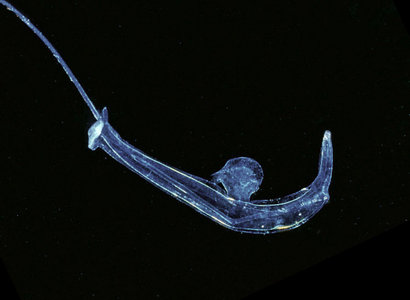
Firoloida is a monotypic genus represented by the species F. desmarestia. It is the smallest (maximal body length of 40 mm) and most transparent species in the family Pterotracheidae. The visceral nucleus is terminal on the long trunk, followed by a very short ventral tail and either a permanent egg string (in females) or a tail filament (in males). Prominent tentacles and a fin sucker are present in males, but are absent in females. The species has a cosmopolitan distribution and is found in tropical to subtropical waters.
Brief Diagnosis
A pterotracheid with:
- Transparent body cylindrical, with a long trunk and a very short tail
- Visceral nucleus terminal on trunk
- Swimming fin anterior of the middle part of the trunk
- Small sucker on anterior margin of swimming fin in males only
- Prominent tentacles ahead of eyes in males only
- Tail filament only in males
- Permanent egg string in females
Characteristics
- Body morphology
- Body elongate, cylindrical
- Trunk long; tail short and ventral
- Visceral nucleus terminal on trunk
- Swimming fin located about one-third the distance back from eyes on the trunk Click on an image to view larger version & data in a new window
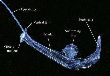
Figure. Body regions and selected structures in a female Firoloida desmarestia. ©
- Sexually dimorphic structures
- Permanent egg string extends posteriorly from base of visceral nucleus in females; length of egg string commonly equal to the body length (Owre, 1964), with early cleavage stages at the proximal end and late-stage veliger larvae at the distal end Click on an image to view larger version & data in a new window
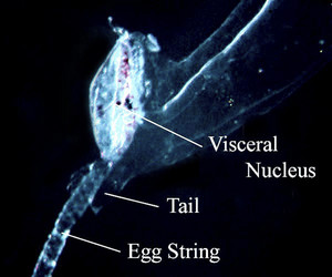
Figure. Posterior body region, right side in a female Firoloida desmarestia. ©
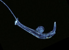
- Filamentous extension of the very short tail in males highly contractile (compare the photograph and drawing below)
- Club-shaped structure (illustrated by Tesch, 1949, but undescribed by any authors to my knowledge) of unknown function. The structure extends posteriorly from the visceral nucleus and consists of a transparent stalk with a white "end bulb" (see photograph below)
Click on an image to view larger version & data in a new window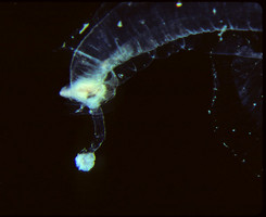
Figure. Posterior portion of body in a male Firoloida desmarestia. ©
- Sucker small and located on anterior margin of swimming fin in males
Click on an image to view larger version & data in a new window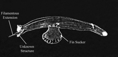
Figure. Drawing of male Firoloida desmarestia, from right side of body. Note the expanded filamentous tail extension. Drawing from Tesch (1949, Fig. 12), modified by addition of labels. © 1949 J. J. Tesch
- Tentacles prominent, located anterior to eyes in males (see photograph below)
- Eye morphology
- Eye shape narrowly triangular
- Lens capsule large and oblong, with lens basal and distal portion slightly opaque
- Larval morphology
- Larva with four velar lobes that are colorless, except for lobe tips that usually have brown spots (Thiriot-Quiévreux, 1973a) Click on an image to view larger version & data in a new window
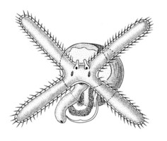
Figure. Drawing of young Firoloida desmarestia larva; shell diameter = 0.3 mm. From Thiriot-Quiévreux (1973a, Fig. 8D; redrawn from Franc, 1948).© 1973 Catherine Thiriot
- Larval shell globular, with about 2 whorls and an aperture that is rounded-ovoid (see SEMs below)
Click on an image to view larger version & data in a new window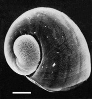
Figure. Scanning electron micrograph of larval shell of Firoloida desmarestia, viewed from right side, angled toward the front of the shell. From Thiriot-Quiévreux (1973a; Fig. 6D). Scale bar - 100 µm. © 1973 Catherine Thiriot
Click on an image to view larger version & data in a new window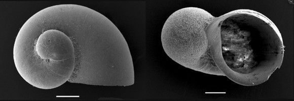
Figure. Scanning electron micrographs of fossil Firoloida desmarestia larval shells from the late Holocene-Recent (Janssen, 2007; Plate 13, Figs. 6a, 4). Left: view of right side (scale bar = 100 µm). Right: apertural view (scale bar = 50 µm). © 2007
- Radula
- Central rachidian tooth consists of a middle portion (with a long and thick median cusp, flanked on either side by three shorter, pointed cusps) and two large, broad-based lateral cusps (Thiriot-Quiévreux, 1973b) Click on an image to view larger version & data in a new window
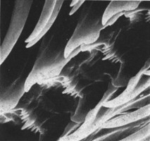
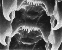
Figure. Scanning electron micrographs of the radula of Firoloida desmarestia. Left: low magnification. Right: high magnification. From Thiriot-Quiévreux (1973b, Plate 1L,K, respectively). © 1973 Catherine Thiriot
- Central rachidian tooth consists of a middle portion (with a long and thick median cusp, flanked on either side by three shorter, pointed cusps) and two large, broad-based lateral cusps (Thiriot-Quiévreux, 1973b)
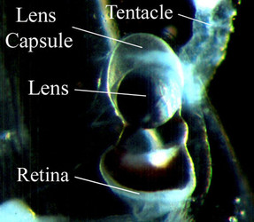
Figure. Dorsal view of right eye in male Firoloida desmarestia. ©
Comments
Ecology: The abundance of Firoloida desmarestia has been reported in very low numbers from the central North Atlantic Ocean (Pafort-van Iersel, 1983), in moderate abundance from the northwestern Indian Ocean (Richter, 1974), and in moderately low to very low numbers from Hawaiian waters of the North Pacific (Seapy, 1990, 2008). Among the heteropods collected by the Meteor Expedition in the Indian Ocean, F. desmarestia ranked ninth (6.1%) of the 22 species collected and accounted for 6.1% of the total numbers (Richter, 1974). In Hawaiian waters it ranked eighth of thirteen heteropod species, representing 4.1% of the total count (Seapy, 1990). And, in a second study (Seapy, 2008) it ranked eleventh of twelve species (0.5% of the total mean nighttime densities) in a fall sampling period and sixteenth of seventeen species (<0.1% of the total mean nighttime densities) in a spring period.
Patterns of vertical distribution in pterotracheids and carinariids were studied by Pafort-van Iersel (1983) based on opening-closing midwater trawl samples collected from the central North Atlantic. No distribution patterns for Firoloida desmarestia could be deduced because only seven specimens were recorded from two tows. In Hawaiian waters of the North Pacific, two opening-closing net studies of diel vertical distribution in heteropods were carried out (Seapy 1990, 2008). In the former paper, F. desmarestia was collected at 0-45, 45-90, and 90-145 m depth intervals during both day and night periods. However, because of high replicate variability among the four samples in each depth interval, there were no statistically significant diel differences in mean abundances, which ranged from 0.5 to 4.5 animals per 1,000 m3. In the latter paper very low numbers of F. desmarestia were collected; it was only recorded in the fall from 80-160 m at the 5 nmi station during the day and at night, and in the spring from 40-60 m at the 15 nmi station during the day.
References
Franc, A. 1948. Véligères et mollusques gastéropodes des Baies d'Alger et de Banyuls. Journal de Conchyliologie (Paris) 88: 3-35, 2 plates.
Janssen, A. W. 2007. Holoplanktonic Mollusca (Gastropoda) from the Gulf of Aqaba, Red Sea and Gulf of Aden (Late Holocene-Recent). Veliger 49: 140-195
Owre, H. B. 1964. Observations on development of the heteropod molluscs Pterotrachea hippocampus and Firoloida desmaresti. Bulletin of Marine Science 14: 519-538.
Pafort-van Iersel, T. 1983. Distribution and variation of Carinariidae and Pterotracheidae (Heteropoda, Gastropoda) of the Amsterdam Mid North Atlantic Plankton Expedition 1980. Beaufortia 33: 73-96.
Richter, G. 1974. Die Heteropoden der "Meteor" Expedition in den Indischen Ozean 1964/65. "Meteor" Forschungs-Ergibnisse Ser. D, No. 17, pp. 55-78.
Seapy, R. R. 1990. Patterns of vertical distribution in epipelagic heteropod molluscs off Hawaii. Marine Ecology Progress Series 60: 235-246.
Seapy, R. R. 2008. Offshore-onshore and vertical distributional patterns of heteropod mollusks off leeward Oahu, Hawaii. Marine Biology 154: 985-995.
Tesch, J. J. 1949. Heteropoda. Dana Report 34, 53 pp., 5 plates.
Thiriot-Quiévreux, C. 1973a. Heteropoda. Oceanography and Marine Biology Annual Review 11:237-261.
Thiriot-Quiévreux, C. 1973b. Observations de la radula des Hétéropodes (Mollusca Prosobranchia) au microscope électronique à balayage et interprétation fonctionnelle. Comptes rendus de l'Académie des Sciences, Serie D 276: 761-764.
Title Illustrations
| Scientific Name | Firoloida desmarestia |
|---|---|
| Location | Monterey Bay, California |
| Specimen Condition | Live Specimen |
| Sex | Female |
| Life Cycle Stage | adult |
| View | left side |
| Copyright | © David Wrobel |
About This Page

California State University, Fullerton, California, USA
Correspondence regarding this page should be directed to Roger R. Seapy at
Page copyright © 2007
 Page: Tree of Life
Firoloida desmarestia .
Authored by
Roger R. Seapy.
The TEXT of this page is licensed under the
Creative Commons Attribution License - Version 3.0. Note that images and other media
featured on this page are each governed by their own license, and they may or may not be available
for reuse. Click on an image or a media link to access the media data window, which provides the
relevant licensing information. For the general terms and conditions of ToL material reuse and
redistribution, please see the Tree of Life Copyright
Policies.
Page: Tree of Life
Firoloida desmarestia .
Authored by
Roger R. Seapy.
The TEXT of this page is licensed under the
Creative Commons Attribution License - Version 3.0. Note that images and other media
featured on this page are each governed by their own license, and they may or may not be available
for reuse. Click on an image or a media link to access the media data window, which provides the
relevant licensing information. For the general terms and conditions of ToL material reuse and
redistribution, please see the Tree of Life Copyright
Policies.
- First online 05 June 2007
- Content changed 08 October 2008
Citing this page:
Seapy, Roger R. 2008. Firoloida desmarestia . Version 08 October 2008. http://tolweb.org/Firoloida_desmarestia/28736/2008.10.08 in The Tree of Life Web Project, http://tolweb.org/


