


Carinaria lamarcki
Roger R. Seapy

Introduction
This is the second largest species (to 220 mm body length) among the six species of Carinaria. The proboscis is short and the trunk moderately long. Both the proboscis and trunk have a thick cutis, the thickness of which increases with age. The tail lacks the thick cutis of the proboscis and trunk. The dorsal crest of the tail is low. The shell is broadly triangular in side view, with the height about one-half the basal length. The keel is moderately low, and increases gradually in height with proximity to the shell aperture.
Brief Diagnosis
A species of Carinaria with:
- Proboscis short and trunk moderately long; both body regions covered with a thick cutis
- Tail lacks the thick cutis of the proboscis and trunk and bears a low dorsal crest
- Shell broadly triangular in side view (height about one-half of the basal length)
- Keel moderately low, increasing gradually in height with proximity to shell aperture
Characteristics
- Body
- Maximal body length 220 mm (Tesch, 1949; from the Mediterranean Sea). In contrast, the largest specimen among the 45 collected by midwater trawls from the middle North Atlantic Ocean (Pafort-van Iersel, 1983) was only 115 mm.
- Proboscis short and trunk moderately long, with both body regions having a thick cutis (see title illustration) that increases in thickness with age (Richter and Seapy, 1999)
- Tail lacks the thick cutis of the proboscis and trunk and has a low dorsal crest (see title illustration)
- Eye shape triangular in dorsal view (click on title illustration for enlarged view of eye)
- Left tentacle long; right one greatly reduced (click on title illustration for enlarged view of left tentacle)
- Maximal body length 220 mm (Tesch, 1949; from the Mediterranean Sea). In contrast, the largest specimen among the 45 collected by midwater trawls from the middle North Atlantic Ocean (Pafort-van Iersel, 1983) was only 115 mm.
- Shell
- Shell broadly triangular in side view (see first photograph below); shell height about one-half the basal length (comparable with C. challengeri)
- Keel moderately well developed; height increases gradually with proximity to shell aperture (see first photograph below)
Click on an image to view larger version & data in a new window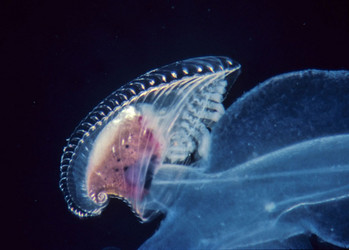
Figure. Stalked visceral nucleus and gills, covered by the transparent shell, in Carinaria lamarcki, viewed from right side of body. © Roger R. Seapy
- Shell apex in adult shells with ventrally-directed protoconch (see first photograph below) Click on an image to view larger version & data in a new window
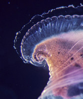
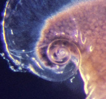
Figure. Apical portion of shell in Carinaria lamarcki, from right side (left) and close up of protoconch (right). © Roger R. Seapy
- Shell spire with two equally-spaced and elevated spiral ridges on the second whorl; the outer ridge ends about one-quarter whorl before the inner one (for an enlarged view, click on the third larval shell SEM below)
- Shell broadly triangular in side view (see first photograph below); shell height about one-half the basal length (comparable with C. challengeri)
- Radula (first SEM below) with central, rachidian teeth (second SEM below) bearing three elongate and pointed median cusps. The median cusps are of unequal length; the middle one is slightly longer than the other two
Click on an image to view larger version & data in a new window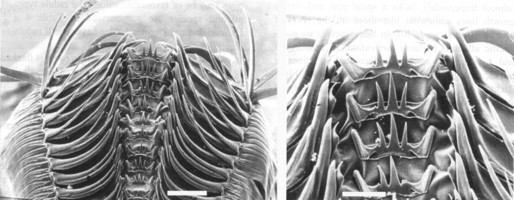
Figure. Scanning electron micrographs of the radula of Carinaria lamarcki at low (left) and high (right) magnifications. Scale bars = 200 µm (left) and 20 µm (right). Photographs from Richter and Seapy (1999). © G. Richter
- Larvae
- Velum comprised of six velar arms; each arm is bordered by a thin line of brown pigment and has a small, terminal brown patch (Thiriot-Quiévreux, 1975)
- Body with brown mantle, digestive tube and opercular lobe; digestive gland yellow (Thiriot-Quiévreux, 1975)
- Larval shell transparent and globular, with an oval aperture and a low, conical spire (see first SEM below). Shell spire (see third SEM below) with two equally-spaced and elevated spiral ridges on the second whorl
Click on an image to view larger version & data in a new windowClick on an image to view larger version & data in a new window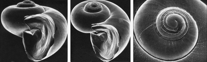
Figure. Scanning electron micrographs of the larval shell of Carinaria lamarcki; apertural view (left), shell tilted about 30° from apertural view (middle), and close up of the spire (right). Photographs from Thiriot-Quiévreux (1975; Fig. 1E-G). © 1975 C. Thiriot
Comments
Only a single study (Pafort-van Iersel, 1983) has been conducted on the diel vertical distribution of Carinaria lamarcki. During the mid North Atlantic Plankton Expedition, paired 1-m2 and 8-m2 opening-closing midwater trawl nets were towed at depth intervals of 50-100 m, at 100 m intervals between 100 and 500 m, and at 500-1,000 m. A total of 45 specimens were collected, which the author regarded as low in comparison with the species' common occurrence in the Mediterranean and Caribbean Seas. Based on this observation, she speculated that C. lamarcki is a neritic species and disperses into the open ocean from the above-mentioned adjacent seas. A noteworthy finding was that no specimens were captured by the 1-m2 net; all were recorded from the 8-m2 net. This result can be explained either as the result of C. lamarcki occurring in such low numbers that the smaller net captured no individuals or, more plausibly, that the species is able to avoid capture by the smaller net. Among the day and night tows from the depth intervals below 200 m sixteen individuals were collected during the day and only one at night (from 300-400 m). In the upper 200 m only one individual was collected during the day (from 100-200 m), while seven were recorded at night (five from 50-100 m and two from 100-200 m). Taken together, these results support an hypothesis of nocturnal vertical migration from mesopelagic into epipelagic waters.
References
Pafort-van Iersel, T. 1983. Distribution and variation of Carinariidae and Pterotracheidae (Heteropoda, Gastropoda) of the Amsterdam Mid North Atlantic Plankton Expedition 1980. Beaufortia 33: 73-96.
Richter, G. and R. R. Seapy. 1999. Heteropoda, pp. 621-647. In: D. Boltovskoy (ed.), South Atlantic Zooplankton. Backhuys Publishers, Leiden.
Seapy, R. R. and C. Thiriot-Quiévreux. 1994. Veliger larvae of Carinariidae (Mollusca: Heteropoda) from Hawaiian waters. Veliger 37: 336-343.
Thiriot-Quiévreux, C. 1975. Observations sur les larves et les adultes de Carinariidae (Mollusca: Heteropoda) de l'Océan Atlantique Nord. Marine Biology 32: 379-388.
Title Illustrations
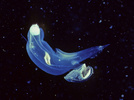
| Scientific Name | Carinaria lamarcki |
|---|---|
| Location | Florida Current |
| Specimen Condition | Live Specimen |
| Identified By | Ronald Gilmer |
| Sex | Female |
| Life Cycle Stage | adult |
| View | right side |
| Copyright | © Ronald Gilmer |
About This Page
Roger R. Seapy

California State University, Fullerton, California, USA
Correspondence regarding this page should be directed to Roger R. Seapy at
rseapy@fullerton.edu
Page copyright © 2008 Roger R. Seapy
 Page: Tree of Life
Carinaria lamarcki .
Authored by
Roger R. Seapy.
The TEXT of this page is licensed under the
Creative Commons Attribution License - Version 3.0. Note that images and other media
featured on this page are each governed by their own license, and they may or may not be available
for reuse. Click on an image or a media link to access the media data window, which provides the
relevant licensing information. For the general terms and conditions of ToL material reuse and
redistribution, please see the Tree of Life Copyright
Policies.
Page: Tree of Life
Carinaria lamarcki .
Authored by
Roger R. Seapy.
The TEXT of this page is licensed under the
Creative Commons Attribution License - Version 3.0. Note that images and other media
featured on this page are each governed by their own license, and they may or may not be available
for reuse. Click on an image or a media link to access the media data window, which provides the
relevant licensing information. For the general terms and conditions of ToL material reuse and
redistribution, please see the Tree of Life Copyright
Policies.
- First online 06 July 2008
- Content changed 23 July 2008
Citing this page:
Seapy, Roger R. 2008. Carinaria lamarcki . Version 23 July 2008 (under construction). http://tolweb.org/Carinaria_lamarcki/28751/2008.07.23 in The Tree of Life Web Project, http://tolweb.org/


