


Abraliopsis sp. A
Lourdes Burgess and Richard E. Young
Introduction
Abraliopsis sp. A is a rather large Abraliopsis reaching at least 46 mm ML. It is known with certainty only from the vicinity of the Hawaiian Islands. It is found throughout much of the central, tropical Pacific.Brief diagnosis:
An Abraliopsis (Pfefferiteuthis) with
- low, short keel and without carpal flap on tentacular club.
- few blue photophores photophore in sectors on anterior mantle.
- a distribution in the tropical Pacific.
Characteristics
In addition to familial characters (listed on the Enoploteuthidae page) and generic characters (listed on the Abraliopsis page), Abraliopsis sp. A has:
- Arms I-III
- Arms I-III with hooks more closely spaced distally. Inner rings of largest suckers with 5 broad teeth distally, smooth proximally.
- Arms I with large trabeculate membranes on both margins. Tubercules trabeculae on distal half of arm; number of tubercules on trabeculae diminishes proximally with none on proximal third of arm. Few tubercules present at base of arm.
- Arms II with trabeculate membrane only on ventral surface. No tubercules on trabeculae; only few tubercules at base of arm.
- Arms III with trabeculate membrane only on ventral surface. Few tubercules present at base of trabeculae in proximal half of arm. Basal region of arm IV with tubercules mostly on lateral margins of oral surface.
- Arms III of males with first basal hook of ventral series enlarged relative to second basal hook of ventral series (see arrows in photograph below). Basal hooks 2-4 of arms III, dorsal series much larger than counterparts of ventral series.
- Females without enlarged hooks on arms I.
Click on an image to view larger version & data in a new window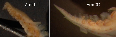
Figure. Oral and oral-oblique views of the arm tip suckers of Abraliopsis sp. A showing suckers in two series. Hawaiian waters, male, 30 mm ML. Photographs by R. Young..
Click on an image to view larger version & data in a new window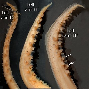
Figure. Oral or dorso-oral views of arms I-III of Abraliopsis sp. A, Hawaiian waters, male, 30 mm ML. Note the relative size of the two basal hooks of arm III, ventral series (arrows). Photograph by R. Young.
- Arms IV (male modifications)
- Right arm IV (hectocotylus):
With large, ventral, rounded-trapezoidal membrane; distally offset smaller dorsal membrane. Small, projecting trabeculae with tubercules present on both borders of proximal arm. Hooks of dorsal series proximally smaller than counterparts of ventral series but become much larger than dorsal counterparts at about hook no. 7.
- Left arm IV:
With greatly enlarged ventral membrane with long trabeculae that project beyond the membrane; trabeculae lined with rows of tuberules (characteristic of subgenus). Smaller projecting trabeculae, with tubercules, present on arm along full dorsal margin. Hooks of dorsal and ventral series about equal is size at arm base, by hook 7/8 ventral hooks distinctly smaller but become equal in size at about hook 17 and may remain so or become smaller near tip . Oral surface of arm at base with tubercules.
Click on an image to view larger version & data in a new window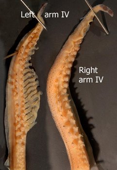
Figure. Oral views of arms IV of Abraliopsis sp. A, Hawaiian waters, male, 30 mm ML. Photograph by R. Young.
- Right arm IV (hectocotylus):
- Tentacles
- Club with carpal flap absent. Keel low, short, on dactylus only.
- Manus with hooks of ventral series nearly 3 times height of counterparts of more dorsal series. Usually 8 hooks present (4D, 4V), rarely 6 or 9.
- Dactylus with with recognizable terminal pad.
- Carpal pad with 4-5 suckers and corresponding knobs.
Click on an image to view larger version & data in a new window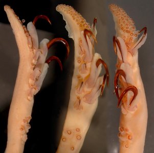
Figure. Dorsal (left), oral (middle) and oral-lateral (right) views of the tentacular club of Abraliopsis sp. A, Hawaiian waters, male, 29 mm ML. Photographs by R. Young.
- Club with carpal flap absent. Keel low, short, on dactylus only.
- Head
- Beaks: Descriptions can be found here: Lower beak; upper beak.
- Occipital crest with occipital folds 3 and 4 joined posteriorly by distinct crescent-shaped ridge.
- Fins
- Fins large, about 70 % of ML.
- Fins large, about 70 % of ML.
- Photophores
- Ocular photophores. 5 ocular photophores with end photophores 2-3 times diameter of middle photophore.
- Integumental photophores: 3 series on ventral head; 6 series on ventral mantle.
Click on an image to view larger version & data in a new window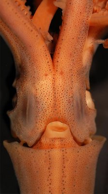
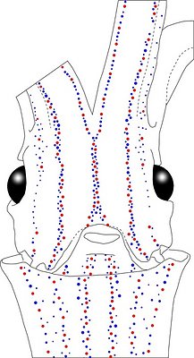
Figure. Ventral views of the integumental photophores of Abraliopsis sp. A, mature male, 30 mm ML, Hawaiian waters. Left - Photograph of the preserved squid. Middle - Outline drawing from photograph with all integumental photophores represented by colored dots. Red dots - Complex photophores. Blue dots - Non-complex photophores. Images by R. Young.
Detailed information on the integumental photophores can be found here.
- Viscera
- Spermatangia receptacles - Females with three pockets for storing spermatangia: One Stellate Pocket at anterior junction of gladius and visceral mass, between stellate ganglia; two Dorsal-collar Pockets on the inner surface of the dorsal part of collar at its anteromedial end.
Click on an image to view larger version & data in a new window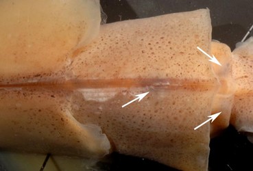
Figure. Dorsal view of the mantle and collar of Abraliopsis sp. A, showing the dark spots (arrows) that mark the sites of deposition of spermatangia; Hawaiian waters, female, 41 mm ML. Photograph by R. Young.
Click on an image to view larger version & data in a new window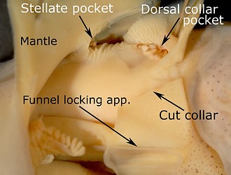
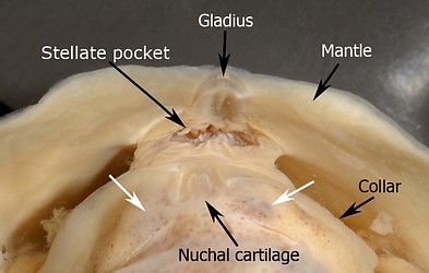
Figure. Spermatangia receptacles (pockets) of Abraliopsis sp. A, Hawaiian waters, females. Left - Side view with both the collar and mantle muscles cut and folded back to show the stellate pocket and the dorsal-collar pocket with a few spermatangia attached; 36 mm ML. Right - Anterior view showing the stellate pocket and spermatangia, and the dark spots (white arrows) that mark the area of spermatangia attachment in the dorsal-collar pockets; 41 mm ML. Photographs by R. Young.
- Spermatophore Click on an image to view larger version & data in a new window
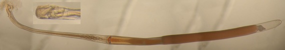
Figure. Spermatophore of Abraliopsis sp. A, Hawaiian waters, male, 37 mm ML. Insert shows the enlarged oral end of another spermatophore from the same squid. Photographs by R. Young.
- Spermatangia receptacles - Females with three pockets for storing spermatangia: One Stellate Pocket at anterior junction of gladius and visceral mass, between stellate ganglia; two Dorsal-collar Pockets on the inner surface of the dorsal part of collar at its anteromedial end.
- Measurements and counts:
TC 32, sta. 37
TC 32, sta. 22
TC 32, sta. 38 TC 32, sta. 56 TC 7, sta. 5
TC 7, sta. 5
TC 48, sta. 16
TC 32, sta. 56
TC 7, sta. 5
Sex
Female
Female
Female
Female
Female
Female
Female
Male
Male
Mantle length
41
39
39
30
35
33
34
34
35
Head width index
29.3
34.3
28.2
35.3
34.3
-
35.3
35.3
28.6
Fin Length index
73.2
-
71.8
73.3
45.7
45.5
67.6
73.5
45.7
Fin width index
102.4
91.4
92.3
93.3
91.4
91
97.1
88.2
97.1
Arm Length index (R/L):
I
53.7/-
-/48.6
41/-
50/-
51.4/-
51.5/-
41.2/-
64.7/-
60/-
II
61/-
-/51.4
59/-
66.7/-
65.7/-
57/6/-
47.1/-
61.8/-
68.6/-
III
53.7/-
-/51.4
51.3+
53.3/-
60
54.5/-
44.1/-
73.5
-/60
IV
65.9/-
-
61.5/61.5
80/70
71.4/80
72.7/66.7 52.9/52.9
73.5/82.4
68.6/71.4
No. arm hooks, R/L; (suckers)
I
22/-; (34)
-
24/-; (10)
20/-; (26)
19/-; (28)
20/-; (28)
20/-; (27)
-
28/-; (24)
II
25/-; (21)
-
25/-; (13)
23/-; (20)
20/-; (19)
26/-; (20)
26/-; (19)
31/-; (19)
31/-; (16)
III
22/-; (18)
21/-; (24)
22/-; -
22/-; (16) 20/-; (14)
22/-; (18)
22/-; (10)
25/-; (13)
30/-; (13)
IV
33/30
33/33
32/28
30/30
31/30
28/47
30/29
32/48
28/43
Club length index (R/L)
26.8/-
31.4/30
51.3/-
26.7/25
31.4/31.4
30.3/30.3
29.4/27.9
29.4/27.9 -
Club hooks, (D/V); (R/L)
4/4; -
3/4; 3/4
4/4; -
4/4; 4/5
4/4; 4/5
4/4; 4/4
4/4; 4/4
4/4; 4/4
-
Carpal suckers (R/L)
5/-
4/4
5/-
5/5
5/5
5/5
6/5
5/5
-
Comments:
Abraliopsis (Pfefferiteuthis) sp. A is very similar to A. falco, Abraliopsis sp. P and A. atlantica. It is separable from A. falco by the
- larger tentacular club (CLI - 30.7 vs 14.2).
- presence of a short keel on the club (keel absent in A. falco).
- greater difference in the relative size of hooks in the dorsal and ventral series of the club (3 X vs. 2 X).
- blue photophores in the Median Mantle Sector more aligned to Sector margins making the central bare strip appear much broader.
It differs from Abraliopsis sp. P in the
- small size of the club keel.
- absence of a carpal flap.
- different shape of the largest club hooks (see "Comments" in A. sp. P description).
- presence of an enlarge first basal hook, ventral series, of arms III in males.
Life History
The development of paralarvae of Abraliopsis sp. A was examined by (Young and Harman, 1985) by hatching eggs collected in the plankton and rearing the hatchlings until all yolk had been utilized (usually 6 days). Older stages were identified by following a morphological series to a size that showed species-specific features.
Eggs are ovoid and about 1.01 by 0.8 mm in major axes, and have a smooth chorion surrounded by a thick layer of jelly and have a small perivitilline space.
Mantle chromatophore pattern of hatchlings is diagnostic (see figure below). Net captured paralarvae generally lose chromatophores due to damage. Terminal arm IV photophores first appear as large swellings at 2.0 mm ML. At 4.6 mm ML, alignment of mantle photophores is apparent. The broad mid-stripe that lacks photophores on the ventral mantle is clearly evident at 8.6 mm ML.
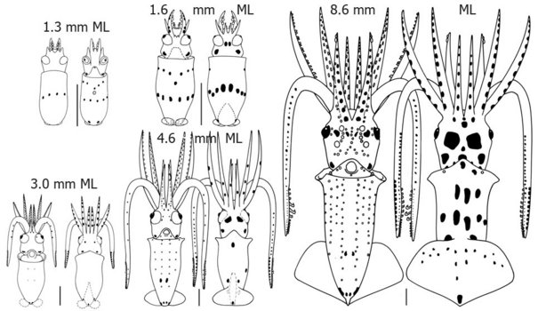
Figure. Ventral and dorsal views of various sizes of paralarvae of Abraliopsis sp. A, Hawaiian waters. The 1.3 mm paralarva is just hatched while the 1.6 mm paralarva is 6 days post-hatching.Drawings from Young and Harman (1985).
Distribution
Geographical distribution: Abraliopsis sp. A is broadly distributed throughout the central tropical Pacific from roughly 25°N to 20°S and from 135°W to 160°W.
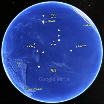
Figure. Distribution of Abraliopsis sp. A. White dots indicate locations where this species has been captured. Base map from Google Earth.app.
Vertical distribution: The vertical distribution was examined in Hawaiian waters (Young, 1978). Squid captured during the day came from about 475-700 m depth with most captures between 550 and 700 m. Nighttime captures were made from depths of about 20-200 m with most squid caught in the upper 100 m. The squid clearly exhibits a diel vertical migration. Paralarvae are captured mostly in the upper 125 m of the water column during both the day and night (Young and Harman, 1985).
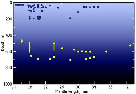
Figure. Vertical distribution chart of Abraliopsis sp A. Captures were made with both open and opening/closing trawls. Bar - fishing depth-range of opening/closing trawl. Dot - Modal fishing depth for either trawl. Blue dot - Night capture. Yellow dot - Day capture. Chart modified from Young (1978, labeled as Abraliopsis sp. A).
References
Young, R. E. 1978. Vertical distribution and photosensitive vesicles of pelagic cephalopods from Hawaiian waters. Fish. Bull., 76: 583-615.
Young, R. E. and R. Harman. 1985. Early life history stages of enoploteuthin squids (Cephalopoda, Teuthoidea, Enoploteuthidae) from Hawaiian waters. Vie et Milieu, 35: 181-202.
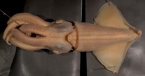
Title Illustrations

| Scientific Name | Abraliopsis sp. A |
|---|---|
| Location | Hawaiian waters |
| Specimen Condition | Preserved |
| Sex | Male |
| Life Cycle Stage | Adult |
| View | Ventral |
| Size | 37 mm ML |
| Image Use |
 This media file is licensed under the Creative Commons Attribution License - Version 3.0. This media file is licensed under the Creative Commons Attribution License - Version 3.0.
|
| Copyright |
©

|
About This Page
Retired
University of Hawaii, Honolulu, HI, USA
Page copyright © 2013 and
 Page: Tree of Life
Abraliopsis sp. A.
Authored by
Lourdes Burgess and Richard E. Young.
The TEXT of this page is licensed under the
Creative Commons Attribution-NonCommercial License - Version 3.0. Note that images and other media
featured on this page are each governed by their own license, and they may or may not be available
for reuse. Click on an image or a media link to access the media data window, which provides the
relevant licensing information. For the general terms and conditions of ToL material reuse and
redistribution, please see the Tree of Life Copyright
Policies.
Page: Tree of Life
Abraliopsis sp. A.
Authored by
Lourdes Burgess and Richard E. Young.
The TEXT of this page is licensed under the
Creative Commons Attribution-NonCommercial License - Version 3.0. Note that images and other media
featured on this page are each governed by their own license, and they may or may not be available
for reuse. Click on an image or a media link to access the media data window, which provides the
relevant licensing information. For the general terms and conditions of ToL material reuse and
redistribution, please see the Tree of Life Copyright
Policies.
- First online 03 November 2013
- Content changed 03 November 2013
Citing this page:
Burgess, Lourdes and Richard E. Young. 2013. Abraliopsis sp. A. Version 03 November 2013 (under construction). http://tolweb.org/Abraliopsis_sp._A/139583/2013.11.03 in The Tree of Life Web Project, http://tolweb.org/


